[English] 日本語
Yorodumi
- EMDB-0148: Structure of alpha-synuclein fibrils -

+
Open data


ID or keywords:

Loading...

-
Basic information

Entry
Database: EMDB / ID: EMD-0148
TitleStructure of alpha-synuclein fibrils
Map dataStructure of alpha-synuclein fibrils
Sample
  • Complex: Alpha-synuclein fibrils
    • Protein or peptide: Alpha-synuclein
Function / homology
Function and homology information


negative regulation of mitochondrial electron transport, NADH to ubiquinone / neutral lipid metabolic process / regulation of phospholipase activity / negative regulation of monooxygenase activity / regulation of acyl-CoA biosynthetic process / negative regulation of dopamine uptake involved in synaptic transmission / negative regulation of norepinephrine uptake / positive regulation of glutathione peroxidase activity / positive regulation of SNARE complex assembly / positive regulation of hydrogen peroxide catabolic process ...negative regulation of mitochondrial electron transport, NADH to ubiquinone / neutral lipid metabolic process / regulation of phospholipase activity / negative regulation of monooxygenase activity / regulation of acyl-CoA biosynthetic process / negative regulation of dopamine uptake involved in synaptic transmission / negative regulation of norepinephrine uptake / positive regulation of glutathione peroxidase activity / positive regulation of SNARE complex assembly / positive regulation of hydrogen peroxide catabolic process / supramolecular fiber / negative regulation of transporter activity / negative regulation of chaperone-mediated autophagy / mitochondrial membrane organization / regulation of reactive oxygen species biosynthetic process / positive regulation of protein localization to cell periphery / regulation of synaptic vesicle recycling / negative regulation of platelet-derived growth factor receptor signaling pathway / negative regulation of exocytosis / regulation of glutamate secretion / response to iron(II) ion / regulation of norepinephrine uptake / dopamine biosynthetic process / SNARE complex assembly / positive regulation of neurotransmitter secretion / synaptic vesicle priming / dopamine uptake involved in synaptic transmission / regulation of macrophage activation / regulation of locomotion / positive regulation of inositol phosphate biosynthetic process / mitochondrial ATP synthesis coupled electron transport / negative regulation of microtubule polymerization / synaptic vesicle transport / dynein complex binding / positive regulation of receptor recycling / regulation of dopamine secretion / positive regulation of endocytosis / protein kinase inhibitor activity / negative regulation of thrombin-activated receptor signaling pathway / response to type II interferon / cuprous ion binding / positive regulation of exocytosis / synaptic vesicle exocytosis / cysteine-type endopeptidase inhibitor activity involved in apoptotic process / response to magnesium ion / kinesin binding / alpha-tubulin binding / regulation of presynapse assembly / synaptic vesicle endocytosis / negative regulation of serotonin uptake / localization / phospholipid metabolic process / supramolecular fiber organization / axon terminus / inclusion body / cellular response to copper ion / Hsp70 protein binding / cellular response to epinephrine stimulus / excitatory postsynaptic potential / response to interleukin-1 / adult locomotory behavior / SNARE binding / positive regulation of release of sequestered calcium ion into cytosol / fatty acid metabolic process / long-term synaptic potentiation / regulation of transmembrane transporter activity / ferrous iron binding / synapse organization / phospholipid binding / protein tetramerization / phosphoprotein binding / microglial cell activation / regulation of long-term neuronal synaptic plasticity / negative regulation of protein kinase activity / tau protein binding / protein destabilization / PKR-mediated signaling / negative regulation of cysteine-type endopeptidase activity involved in apoptotic process / positive regulation of protein serine/threonine kinase activity / receptor internalization / synaptic vesicle membrane / positive regulation of inflammatory response / activation of cysteine-type endopeptidase activity involved in apoptotic process / actin cytoskeleton / positive regulation of peptidyl-serine phosphorylation / cellular response to oxidative stress / actin binding / cell cortex / histone binding / growth cone / chemical synaptic transmission / postsynapse / neuron apoptotic process / negative regulation of neuron apoptotic process / amyloid fibril formation / response to lipopolysaccharide / lysosome / oxidoreductase activity / molecular adaptor activity / transcription cis-regulatory region binding
Similarity search - Function
Synuclein / Alpha-synuclein / Synuclein
Similarity search - Domain/homology
Biological speciesHomo sapiens (human)
Methodhelical reconstruction / cryo EM / Resolution: 3.4 Å
AuthorsGuerrero-Ferreira R / Taylor NMI / Mona D / Ringler P / Lauer ME / Riek R / Britschgi M / Stahlberg H
Citation
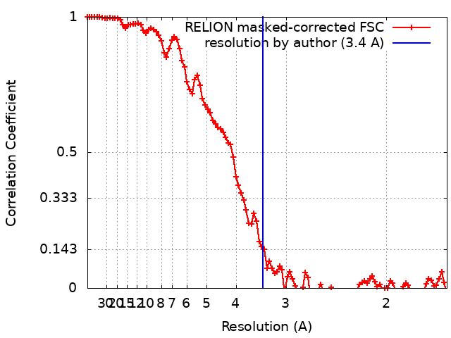
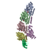
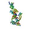
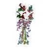
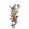
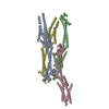
Journal: Elife / Year: 2018
Title: Cryo-EM structure of alpha-synuclein fibrils.
Authors: Ricardo Guerrero-Ferreira / Nicholas Mi Taylor / Daniel Mona / Philippe Ringler / Matthias E Lauer / Roland Riek / Markus Britschgi / Henning Stahlberg /
Abstract: Parkinson's disease is a progressive neuropathological disorder that belongs to the class of synucleinopathies, in which the protein alpha-synuclein is found at abnormally high concentrations in ...Parkinson's disease is a progressive neuropathological disorder that belongs to the class of synucleinopathies, in which the protein alpha-synuclein is found at abnormally high concentrations in affected neurons. Its hallmark are intracellular inclusions called Lewy bodies and Lewy neurites. We here report the structure of cytotoxic alpha-synuclein fibrils (residues 1-121), determined by cryo-electron microscopy at a resolution of 3.4 Å. Two protofilaments form a polar fibril composed of staggered β-strands. The backbone of residues 38 to 95, including the fibril core and the non-amyloid component region, are well resolved in the EM map. Residues 50-57, containing three of the mutation sites associated with familial synucleinopathies, form the interface between the two protofilaments and contribute to fibril stability. A hydrophobic cleft at one end of the fibril may have implications for fibril elongation, and invites for the design of molecules for diagnosis and treatment of synucleinopathies.
History
Header (metadata) releaseMar 14, 2018-
DepositionJul 26, 2018-
Map releaseAug 8, 2018-
SupersessionFeb 15, 2023ID: EMD-4276
UpdateFeb 22, 2023-
Current statusFeb 22, 2023Processing site: PDBe / Status: Released

-
Structure visualization
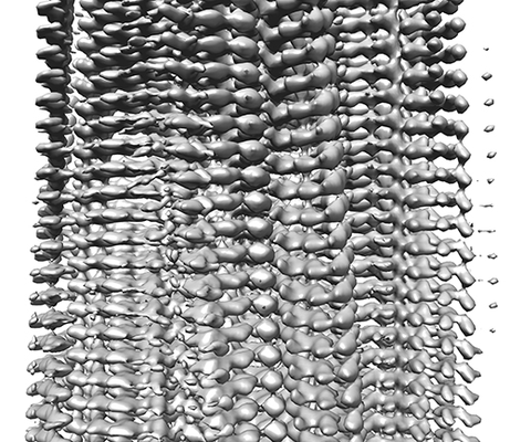
Movie
  • Surface view with section colored by density value
  • Surface level: 0.029
  • Imaged by UCSF Chimera
  • Download
  • Surface view colored by cylindrical radius
  • Surface level: 0.029
  • Imaged by UCSF Chimera
  • Download
  • Surface view with fitted model
  • Atomic models: PDB-6h6b
  • Surface level: 0.029
  • Imaged by UCSF Chimera
  • Download
  • Simplified surface model + fitted atomic model
  • Atomic modelsPDB-6h6b
  • Imaged by Jmol
  • Download
Movie viewer
Structure viewerEM map:
SurfViewMolmilJmol/JSmol
Supplemental images

Downloads & links

-
Map

FileDownload / File: emd_0148.map.gz / Format: CCP4 / Size: 83.7 MB / Type: IMAGE STORED AS FLOATING POINT NUMBER (4 BYTES)
AnnotationStructure of alpha-synuclein fibrils
Voxel sizeX=Y=Z: 0.831 Å
Density
Contour LevelBy AUTHOR: 0.029 / Movie #1: 0.029
Minimum - Maximum-0.08086198 - 0.16965547
Average (Standard dev.)0.0020613861 (±0.007307958)
SymmetrySpace group: 1
Details

EMDB XML:

Map geometry
Axis orderXYZ
Origin000
Dimensions280280280
Spacing280280280
CellA=B=C: 232.68 Å
α=β=γ: 90.0 °

CCP4 map header:

modeImage stored as Reals
Å/pix. X/Y/Z0.8310.8310.831
M x/y/z280280280
origin x/y/z0.0000.0000.000
length x/y/z232.680232.680232.680
α/β/γ90.00090.00090.000
MAP C/R/S123
start NC/NR/NS000
NC/NR/NS280280280
D min/max/mean-0.0810.1700.002

-
Supplemental data

-
Sample components

-
Entire : Alpha-synuclein fibrils

EntireName: Alpha-synuclein fibrils
Components
  • Complex: Alpha-synuclein fibrils
    • Protein or peptide: Alpha-synuclein

-
Supramolecule #1: Alpha-synuclein fibrils

SupramoleculeName: Alpha-synuclein fibrils / type: complex / ID: 1 / Parent: 0 / Macromolecule list: all / Details: Residues 1-121
Source (natural)Organism: Homo sapiens (human)

-
Macromolecule #1: Alpha-synuclein

MacromoleculeName: Alpha-synuclein / type: protein_or_peptide / ID: 1 / Number of copies: 10 / Enantiomer: LEVO
Source (natural)Organism: Homo sapiens (human)
Molecular weightTheoretical: 12.242873 KDa
Recombinant expressionOrganism: Escherichia coli 'BL21-Gold(DE3)pLysS AG' (bacteria)
SequenceString:
MDVFMKGLSK AKEGVVAAAE KTKQGVAEAA GKTKEGVLYV GSKTKEGVVH GVATVAEKTK EQVTNVGGAV VTGVTAVAQK TVEGAGSIA AATGFVKKDQ LGKNEEGAPQ EGILEDMPVD PD

-
Experimental details

-
Structure determination

Methodcryo EM
Processinghelical reconstruction
Aggregation statefilament

-
Sample preparation

Concentration5 mg/mL
BufferpH: 7.3
Component:
ConcentrationFormulaName
2.66 mMKClPotassium chloride
1.47 mMKH2PO4Potassium phosphate
137.93 mMNaClSodium chlorideSodium chloride
8.06 mMNa2HPO4Sodium phosphate
GridModel: Quantifoil R2/2 / Material: COPPER / Mesh: 300 / Support film - Material: CARBON / Support film - topology: HOLEY / Pretreatment - Type: GLOW DISCHARGE / Pretreatment - Time: 20 sec.
VitrificationCryogen name: ETHANE / Chamber humidity: 95 % / Chamber temperature: 277 K / Instrument: FEI VITROBOT MARK IV

-
Electron microscopy

MicroscopeFEI TITAN KRIOS
Electron beamAcceleration voltage: 300 kV / Electron source: FIELD EMISSION GUN
Electron opticsIllumination mode: FLOOD BEAM / Imaging mode: BRIGHT FIELDBright-field microscopy
Specialist opticsEnergy filter - Name: GIF Quantum LS
Image recordingFilm or detector model: GATAN K2 SUMMIT (4k x 4k) / Detector mode: COUNTING / Digitization - Frames/image: 2-50 / Average electron dose: 100.0 e/Å2
Experimental equipment
Model: Titan Krios / Image courtesy: FEI Company

-
Image processing

Segment selectionNumber selected: 18860 / Software - Name: RELION
Details: Segments extracted from 792 manually picked fibrils
Final angle assignmentType: PROJECTION MATCHING / Software - Name: RELION
Final reconstructionApplied symmetry - Helical parameters - Δz: 2.45 Å
Applied symmetry - Helical parameters - Δ&Phi: 179.5 °
Applied symmetry - Helical parameters - Axial symmetry: C1 (asymmetric)
Resolution.type: BY AUTHOR / Resolution: 3.4 Å / Resolution method: FSC 0.143 CUT-OFF / Software - Name: RELION / Number images used: 13390
FSC plot (resolution estimation)

+
About Yorodumi

-
News

-
Feb 9, 2022. New format data for meta-information of EMDB entries

New format data for meta-information of EMDB entries

  • Version 3 of the EMDB header file is now the official format.
  • The previous official version 1.9 will be removed from the archive.

Related info.:EMDB header

External links:wwPDB to switch to version 3 of the EMDB data model

-
Aug 12, 2020. Covid-19 info

Covid-19 info

URL: https://pdbj.org/emnavi/covid19.php

New page: Covid-19 featured information page in EM Navigator.

Related info.:Covid-19 info / Mar 5, 2020. Novel coronavirus structure data

+
Mar 5, 2020. Novel coronavirus structure data

Novel coronavirus structure data

Related info.:Yorodumi Speices / Aug 12, 2020. Covid-19 info

External links:COVID-19 featured content - PDBj / Molecule of the Month (242):Coronavirus Proteases

+
Jan 31, 2019. EMDB accession codes are about to change! (news from PDBe EMDB page)

EMDB accession codes are about to change! (news from PDBe EMDB page)

  • The allocation of 4 digits for EMDB accession codes will soon come to an end. Whilst these codes will remain in use, new EMDB accession codes will include an additional digit and will expand incrementally as the available range of codes is exhausted. The current 4-digit format prefixed with “EMD-” (i.e. EMD-XXXX) will advance to a 5-digit format (i.e. EMD-XXXXX), and so on. It is currently estimated that the 4-digit codes will be depleted around Spring 2019, at which point the 5-digit format will come into force.
  • The EM Navigator/Yorodumi systems omit the EMD- prefix.

Related info.:Q: What is EMD? / ID/Accession-code notation in Yorodumi/EM Navigator

External links:EMDB Accession Codes are Changing Soon! / Contact to PDBj

+
Jul 12, 2017. Major update of PDB

Major update of PDB

  • wwPDB released updated PDB data conforming to the new PDBx/mmCIF dictionary.
  • This is a major update changing the version number from 4 to 5, and with Remediation, in which all the entries are updated.
  • In this update, many items about electron microscopy experimental information are reorganized (e.g. em_software).
  • Now, EM Navigator and Yorodumi are based on the updated data.

External links:wwPDB Remediation / Enriched Model Files Conforming to OneDep Data Standards Now Available in the PDB FTP Archive

-
Yorodumi

Thousand views of thousand structures

  • Yorodumi is a browser for structure data from EMDB, PDB, SASBDB, etc.
  • This page is also the successor to EM Navigator detail page, and also detail information page/front-end page for Omokage search.
  • The word "yorodu" (or yorozu) is an old Japanese word meaning "ten thousand". "mi" (miru) is to see.

Related info.:EMDB / PDB / SASBDB / Comparison of 3 databanks / Yorodumi Search / Aug 31, 2016. New EM Navigator & Yorodumi / Yorodumi Papers / Jmol/JSmol / Function and homology information / Changes in new EM Navigator and Yorodumi

Read more