[English] 日本語
Yorodumi Papers
- Database of articles cited by EMDB/PDB/SASBDB data -

+
Search query

Keywords
Structure methods
Author
Journal
IF

-
Structure paper

TitleStructure of the SARS-CoV-2 main protease
Journal, issue, pagesTo Be Published
Publish dateMay 10, 2024 (structure data deposition date)
AuthorsBlankenship, L.R. / Liu, W.R.
External linksSearch PubMed
MethodsX-ray diffraction
Resolution1.6 - 2.5 Å
Structure data

PDB-9bql:
Structure of the SARS-CoV-2 main protease in complex with inhibitor VB-A-32
Method: X-RAY DIFFRACTION / Resolution: 1.8 Å

PDB-9bqm:
Structure of the SARS-CoV-2 main protease in complex with inhibitor VB-A-26
Method: X-RAY DIFFRACTION / Resolution: 2.1 Å

PDB-9bqo:
Structure of the SARS-CoV-2 main protease in complex with inhibitor k88
Method: X-RAY DIFFRACTION / Resolution: 2.5 Å

PDB-9bqp:
Structure of the SARS-CoV-2 main protease in complex with inhibitor R79
Method: X-RAY DIFFRACTION / Resolution: 2 Å

PDB-9bqq:
Structure of the SARS-CoV-2 main protease in complex with inhibitor R81
Method: X-RAY DIFFRACTION / Resolution: 2 Å

PDB-9bqt:
Structure of the SARS-CoV-2 main protease in complex with inhibitor R80
Method: X-RAY DIFFRACTION / Resolution: 2.1 Å

PDB-9bqy:
Structure of the SARS-CoV-2 main protease in complex with inhibitor R70
Method: X-RAY DIFFRACTION / Resolution: 2.5 Å

PDB-9bqz:
Structure of the SARS-CoV-2 main protease in complex with inhibitor x11
Method: X-RAY DIFFRACTION / Resolution: 1.7 Å

PDB-9br0:
Structure of the SARS-CoV-2 main protease in complex with inhibitor YR-B-84
Method: X-RAY DIFFRACTION / Resolution: 2.5 Å

PDB-9br1:
Structure of the SARS-CoV-2 main protease in complex with inhibitor VB-A-70
Method: X-RAY DIFFRACTION / Resolution: 1.9 Å

PDB-9bs7:
Structure of the SARS-CoV-2 main protease in complex with inhibitor Vinylpyridine
Method: X-RAY DIFFRACTION / Resolution: 1.85 Å

PDB-9bs8:
Structure of the SARS-CoV-2 main protease in complex with inhibitor YR-B-107
Method: X-RAY DIFFRACTION / Resolution: 1.7 Å

PDB-9bsa:
Structure of the SARS-CoV-2 main protease in complex with inhibitor VB-B-112
Method: X-RAY DIFFRACTION / Resolution: 1.7 Å

PDB-9bse:
Structure of the SARS-CoV-2 main protease in complex with inhibitor YR-B-165
Method: X-RAY DIFFRACTION / Resolution: 1.85 Å

PDB-9bsf:
Structure of the SARS-CoV-2 main protease in complex with inhibitor SR-A-171
Method: X-RAY DIFFRACTION / Resolution: 1.75 Å

PDB-9bsg:
Structure of the SARS-CoV-2 main protease in complex with inhibitor VB-C-20
Method: X-RAY DIFFRACTION / Resolution: 1.75 Å

PDB-9bsi:
Structure of the SARS-CoV-2 main protease in complex with inhibitor SR-B-7
Method: X-RAY DIFFRACTION / Resolution: 1.7 Å

PDB-9bso:
Structure of the SARS-CoV-2 main protease in complex with inhibitor SR-B-13
Method: X-RAY DIFFRACTION / Resolution: 1.7 Å

PDB-9bsp:
Structure of the SARS-CoV-2 main protease in complex with inhibitor VB-C-68
Method: X-RAY DIFFRACTION / Resolution: 1.6 Å

PDB-9bsq:
Structure of the SARS-CoV-2 main protease in complex with inhibitor VB-C-70
Method: X-RAY DIFFRACTION / Resolution: 1.8 Å

PDB-9bsr:
Structure of the SARS-CoV-2 main protease in complex with inhibitor YR-B-136B
Method: X-RAY DIFFRACTION / Resolution: 1.9 Å

PDB-9bst:
Structure of the SARS-CoV-2 main protease in complex with inhibitor CID8009_5647
Method: X-RAY DIFFRACTION / Resolution: 1.6 Å

PDB-9bte:
Structure of the SARS-CoV-2 main protease in complex with inhibitor CID5573_0017
Method: X-RAY DIFFRACTION / Resolution: 1.6 Å

PDB-9btf:
Structure of the SARS-CoV-2 main protease in complex with inhibitor SR-B-77
Method: X-RAY DIFFRACTION / Resolution: 1.8 Å

PDB-9btk:
Structure of the SARS-CoV-2 main protease in complex with inhibitor YR-C-108T
Method: X-RAY DIFFRACTION / Resolution: 1.79 Å

PDB-9btr:
Structure of the SARS-CoV-2 main protease in complex with inhibitor YR-C-163
Method: X-RAY DIFFRACTION / Resolution: 1.9 Å

PDB-9btt:
Structure of the SARS-CoV-2 main protease in complex with inhibitor SR-B-51T
Method: X-RAY DIFFRACTION / Resolution: 1.8 Å

Chemicals

PDB-1arg:
Aspartate aminotransferase, phospho-5'-pyridoxyl aspartate complex

ChemComp-HOH:
WATER

PDB-1ari:
Aspartate aminotransferase, W140H mutant, maleate complex

PDB-1arm:
CARBOXYPEPTIDASE A WITH ZN REPLACED BY HG

PDB-1arh:
ASPARTATE AMINOTRANSFERASE, Y225R/R386A MUTANT

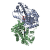
PDB-1ars:
X-RAY CRYSTALLOGRAPHIC STUDY OF PYRIDOXAL 5'-PHOSPHATE-TYPE ASPARTATE AMINOTRANSFERASES FROM ESCHERICHIA COLI IN OPEN AND CLOSED FORM

PDB-1art:
X-RAY CRYSTALLOGRAPHIC STUDY OF PYRIDOXAL 5'-PHOSPHATE-TYPE ASPARTATE AMINOTRANSFERASES FROM ESCHERICHIA COLI IN OPEN AND CLOSED FORM

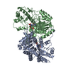
PDB-1arv:
CRYSTAL STRUCTURES OF CYANIDE-AND TRIIODIDE-BOUND FORMS OF ARTHROMYCES RAMOSUS PEROXIDASE AT DIFFERENT PH VALUES. PERTURBATIONS OF ACTIVE SITE RESIDUES AND THEIR IMPLICATION IN ENZYME CATALYSIS

PDB-1aru:
CRYSTAL STRUCTURES OF CYANIDE-AND TRIIODIDE-BOUND FORMS OF ARTHROMYCES RAMOSUS PEROXIDASE AT DIFFERENT PH VALUES. PERTURBATIONS OF ACTIVE SITE RESIDUES AND THEIR IMPLICATION IN ENZYME CATALYSIS

PDB-1arr:
RELAXATION MATRIX REFINEMENT OF THE SOLUTION STRUCTURE OF THE ARC REPRESSOR

PDB-1ar4:
X-RAY STRUCTURE ANALYSIS OF THE CAMBIALISTIC SUPEROXIDE DISMUTASE FROM PROPIONIBACTERIUM SHERMANII ACTIVE WITH FE OR MN

PDB-1ar5:
X-RAY STRUCTURE OF THE CAMBIALISTIC SUPEROXIDE DISMUTASE FROM PROPIONIBACTERIUM SHERMANII ACTIVE WITH FE OR MN

PDB-1ar6:
P1/MAHONEY POLIOVIRUS, DOUBLE MUTANT V1160I +P1095S

PDB-1ar7:
P1/MAHONEY POLIOVIRUS, DOUBLE MUTANT P1095S + H2142Y

PDB-1ar8:
P1/MAHONEY POLIOVIRUS, MUTANT P1095S

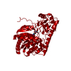
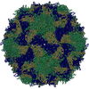
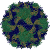
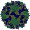
PDB-1ar9:
P1/MAHONEY POLIOVIRUS, SINGLE SITE MUTANT H2142Y

PDB-1arw:
CRYSTAL STRUCTURES OF CYANIDE-AND TRIIODIDE-BOUND FORMS OF ARTHROMYCES RAMOSUS PEROXIDASE AT DIFFERENT PH VALUES. PERTURBATIONS OF ACTIVE SITE RESIDUES AND THEIR IMPLICATION IN ENZYME CATALYSIS

PDB-1arx:
CRYSTAL STRUCTURES OF CYANIDE-AND TRIIODIDE-BOUND FORMS OF ARTHROMYCES RAMOSUS PEROXIDASE AT DIFFERENT PH VALUES. PERTURBATIONS OF ACTIVE SITE RESIDUES AND THEIR IMPLICATION IN ENZYME CATALYSIS

PDB-1ary:
CRYSTAL STRUCTURES OF CYANIDE-AND TRIIODIDE-BOUND FORMS OF ARTHROMYCES RAMOSUS PEROXIDASE AT DIFFERENT PH VALUES. PERTURBATIONS OF ACTIVE SITE RESIDUES AND THEIR IMPLICATION IN ENZYME CATALYSIS

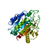
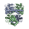
PDB-1asa:
THE STRUCTURAL BASIS FOR THE REDUCED ACTIVITY OF THE Y226F(Y225F) ACTIVE SITE MUTANT OF E. COLI ASPARTATE AMINOTRANSFERASE

ChemComp-A5Z:
[3-[2,6-bis(chloranyl)phenyl]-5-methyl-1,2-oxazol-4-yl]methanol

PDB-1asf:
THE STRUCTURAL BASIS FOR THE REDUCED ACTIVITY OF THE Y226F(Y225F) ACTIVE SITE MUTANT OF E. COLI ASPARTATE AMINOTRANSFERASE

PDB-1ash:
THE STRUCTURE OF ASCARIS HEMOGLOBIN DOMAIN I AT 2.2 ANGSTROMS RESOLUTION: MOLECULAR FEATURES OF OXYGEN AVIDITY

PDB-1asg:
THE STRUCTURAL BASIS FOR THE REDUCED ACTIVITY OF THE Y226F(Y225F) ACTIVE SITE MUTANT OF E. COLI ASPARTATE AMINOTRANSFERASE

PDB-1asj:
P1/MAHONEY POLIOVIRUS, AT CRYOGENIC TEMPERATURE

Source
  • severe acute respiratory syndrome coronavirus 2
KeywordsVIRAL PROTEIN / Hydrolase / main protease

+
About Yorodumi Papers

-
News

-
Feb 9, 2022. New format data for meta-information of EMDB entries

New format data for meta-information of EMDB entries

  • Version 3 of the EMDB header file is now the official format.
  • The previous official version 1.9 will be removed from the archive.

Related info.:EMDB header

External links:wwPDB to switch to version 3 of the EMDB data model

-
Aug 12, 2020. Covid-19 info

Covid-19 info

URL: https://pdbj.org/emnavi/covid19.php

New page: Covid-19 featured information page in EM Navigator.

Related info.:Covid-19 info / Mar 5, 2020. Novel coronavirus structure data

+
Mar 5, 2020. Novel coronavirus structure data

Novel coronavirus structure data

Related info.:Yorodumi Speices / Aug 12, 2020. Covid-19 info

External links:COVID-19 featured content - PDBj / Molecule of the Month (242):Coronavirus Proteases

+
Jan 31, 2019. EMDB accession codes are about to change! (news from PDBe EMDB page)

EMDB accession codes are about to change! (news from PDBe EMDB page)

  • The allocation of 4 digits for EMDB accession codes will soon come to an end. Whilst these codes will remain in use, new EMDB accession codes will include an additional digit and will expand incrementally as the available range of codes is exhausted. The current 4-digit format prefixed with “EMD-” (i.e. EMD-XXXX) will advance to a 5-digit format (i.e. EMD-XXXXX), and so on. It is currently estimated that the 4-digit codes will be depleted around Spring 2019, at which point the 5-digit format will come into force.
  • The EM Navigator/Yorodumi systems omit the EMD- prefix.

Related info.:Q: What is EMD? / ID/Accession-code notation in Yorodumi/EM Navigator

External links:EMDB Accession Codes are Changing Soon! / Contact to PDBj

+
Jul 12, 2017. Major update of PDB

Major update of PDB

  • wwPDB released updated PDB data conforming to the new PDBx/mmCIF dictionary.
  • This is a major update changing the version number from 4 to 5, and with Remediation, in which all the entries are updated.
  • In this update, many items about electron microscopy experimental information are reorganized (e.g. em_software).
  • Now, EM Navigator and Yorodumi are based on the updated data.

External links:wwPDB Remediation / Enriched Model Files Conforming to OneDep Data Standards Now Available in the PDB FTP Archive

-
Yorodumi Papers

Database of articles cited by EMDB/PDB/SASBDB data

  • Database of articles cited by EMDB, PDB, and SASBDB entries
  • Using PubMed data

Related info.:EMDB / PDB / SASBDB / Yorodumi / EMN Papers / Changes in new EM Navigator and Yorodumi

Read more