 Movie
Movie Controller
 Structure viewers
 About Yorodumi Papers


+Search query
-Structure paper
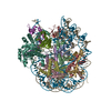
| Title | Basis of the H2AK119 specificity of the Polycomb repressive deubiquitinase. |
|---|---|
| Journal, issue, pages | Nature, Vol. 616, Issue 7955, Page 176-182, Year 2023 |
| Publish date | Mar 29, 2023 |
 Authors Authors | Weiran Ge / Cong Yu / Jingjing Li / Zhenyu Yu / Xiaorong Li / Yan Zhang / Chao-Pei Liu / Yingfeng Li / Changlin Tian / Xinzheng Zhang / Guohong Li / Bing Zhu / Rui-Ming Xu /  |
| PubMed Abstract | Repression of gene expression by protein complexes of the Polycomb group is a fundamental mechanism that governs embryonic development and cell-type specification. The Polycomb repressive ...Repression of gene expression by protein complexes of the Polycomb group is a fundamental mechanism that governs embryonic development and cell-type specification. The Polycomb repressive deubiquitinase (PR-DUB) complex removes the ubiquitin moiety from monoubiquitinated histone H2A K119 (H2AK119ub1) on the nucleosome, counteracting the ubiquitin E3 ligase activity of Polycomb repressive complex 1 (PRC1) to facilitate the correct silencing of genes by Polycomb proteins and safeguard active genes from inadvertent silencing by PRC1 (refs. ). The intricate biological function of PR-DUB requires accurate targeting of H2AK119ub1, but PR-DUB can deubiquitinate monoubiquitinated free histones and peptide substrates indiscriminately; the basis for its exquisite nucleosome-dependent substrate specificity therefore remains unclear. Here we report the cryo-electron microscopy structure of human PR-DUB, composed of BAP1 and ASXL1, in complex with the chromatosome. We find that ASXL1 directs the binding of the positively charged C-terminal extension of BAP1 to nucleosomal DNA and histones H3-H4 near the dyad, an addition to its role in forming the ubiquitin-binding cleft. Furthermore, a conserved loop segment of the catalytic domain of BAP1 is situated near the H2A-H2B acidic patch. This distinct nucleosome-binding mode displaces the C-terminal tail of H2A from the nucleosome surface, and endows PR-DUB with the specificity for H2AK119ub1. |
 External links External links | Nature / PubMed:36991118 |
| Methods | EM (single particle) |
| Resolution | 2.75 - 4.5 Å |
| Structure data | EMDB-34431, PDB-8h1t:  EMDB-34432: Cryo-EM structure of BAP1-ASXL1 bound to nucleosome  EMDB-35179: Constituent map of cryo-EM structure of BAP1-ASXL1 bound to chromatosome  EMDB-35180: Constituent map of cryo-EM structure of BAP1-ASXL1 bound to chromatosome  EMDB-35181: Constituent map of cryo-EM structure of BAP1-ASXL1 bound to chromatosome  EMDB-35182: Consensus map of cryo-EM structure of BAP1-ASXL1 bound to chromatosome |
| Source |
|
 Keywords Keywords | GENE REGULATION / nucleosome / histone deubiquitination / chromatin |

 homo sapiens (human)