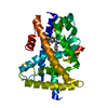
PDB-6kax: X-ray structure of human PPARalpha ligand binding domain-intrinsic fatty acid (E. coli origin) co-crystals obtained by cross-seeding 手法: X-RAY DIFFRACTION / 解像度: 1.23 Å
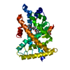
PDB-6kay: X-ray structure of human PPARalpha ligand binding domain-GW7647 co-crystals obtained by soaking 手法: X-RAY DIFFRACTION / 解像度: 1.735 Å
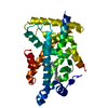
PDB-6kaz: X-ray structure of human PPARalpha ligand binding domain-pemafibrate co-crystals obtained by soaking 手法: X-RAY DIFFRACTION / 解像度: 1.48 Å
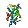
PDB-6kb0: X-ray structure of human PPARalpha ligand binding domain-5,8,11,14-eicosatetraynoic acid (ETYA) co-crystals obtained by soaking 手法: X-RAY DIFFRACTION / 解像度: 1.35 Å
PDB-6kb1: X-ray structure of human PPARalpha ligand binding domain-tetradecylthioacetic acid (TTA) co-crystals obtained by soaking 手法: X-RAY DIFFRACTION / 解像度: 1.25 Å
PDB-6kb2: X-ray structure of human PPARalpha ligand binding domain-Wy14643 co-crystals obtained by soaking 手法: X-RAY DIFFRACTION / 解像度: 1.95 Å
PDB-6kb3: X-ray structure of human PPARalpha ligand binding domain-GW7647 co-crystals obtained by delipidation and cross-seeding 手法: X-RAY DIFFRACTION / 解像度: 1.45 Å
PDB-6kb4: X-ray structure of human PPARalpha ligand binding domain-pemafibrate co-crystals obtained by delipidation and cross-seeding 手法: X-RAY DIFFRACTION / 解像度: 1.42 Å
PDB-6kb5: X-ray structure of human PPARalpha ligand binding domain-5,8,11,14-eicosatetraynoic Acid (ETYA) co-crystals obtained by delipidation and cross-seeding 手法: X-RAY DIFFRACTION / 解像度: 1.95 Å
PDB-6kb6: X-ray structure of human PPARalpha ligand binding domain-tetradecylthioacetic acid (TTA) co-crystals obtained by delipidation and cross-seeding 手法: X-RAY DIFFRACTION / 解像度: 1.431 Å
PDB-6kb7: X-ray structure of human PPARalpha ligand binding domain-Wy14643 co-crystals obtained by delipidation and cross-seeding 手法: X-RAY DIFFRACTION / 解像度: 2.14 Å
PDB-6kb8: X-ray structure of human PPARalpha ligand binding domain-GW7647 co-crystals obtained by cross-seeding 手法: X-RAY DIFFRACTION / 解像度: 1.47 Å
PDB-6kb9: X-ray structure of human PPARalpha ligand binding domain-pemafibrate co-crystals obtained by cross-seeding 手法: X-RAY DIFFRACTION / 解像度: 1.55 Å
PDB-6kba: X-ray structure of human PPARalpha ligand binding domain-Wy14643 co-crystals obtained by co-crystallization 手法: X-RAY DIFFRACTION / 解像度: 1.82 Å
PDB-6kyp: X-ray structure of human PPARalpha ligand binding domain-GW9662-clofibric acid co-crystals obtained by delipidation and co-crystallization 手法: X-RAY DIFFRACTION / 解像度: 2.86 Å
PDB-6l36: X-ray structure of human PPARalpha ligand binding domain-GW9662-fenofibric acid co-crystals obtained by delipidation and co-crystallization 手法: X-RAY DIFFRACTION / 解像度: 3.301 Å
PDB-6l37: X-ray structure of human PPARalpha ligand binding domain-GW9662-ciprofibrate co-crystals obtained by delipidation and co-crystallization 手法: X-RAY DIFFRACTION / 解像度: 2.91 Å
PDB-6l38: X-ray structure of human PPARalpha ligand binding domain-GW9662-gemfibrozil co-crystals obtained by delipidation and co-crystallization 手法: X-RAY DIFFRACTION / 解像度: 2.761 Å
PDB-6lx4: X-ray structure of human PPARalpha ligand binding domain-fenofibric acid co-crystals obtained by delipidation and co-crystallization 手法: X-RAY DIFFRACTION / 解像度: 2.13 Å
PDB-6lx5: X-ray structure of human PPARalpha ligand binding domain-ciprofibrate co-crystals obtained by delipidation and co-crystallization 手法: X-RAY DIFFRACTION / 解像度: 1.87 Å
PDB-6lx6: X-ray structure of human PPARalpha ligand binding domain-palmitic acid co-crystals obtained by delipidation and cross-seeding 手法: X-RAY DIFFRACTION / 解像度: 1.3 Å
PDB-6lx7: X-ray structure of human PPARalpha ligand binding domain-stearic acid co-crystals obtained by delipidation and cross-seeding 手法: X-RAY DIFFRACTION / 解像度: 1.41 Å
PDB-6lx8: X-ray structure of human PPARalpha ligand binding domain-oleic acid co-crystals obtained by delipidation and cross-seeding 手法: X-RAY DIFFRACTION / 解像度: 1.54 Å
PDB-6lx9: X-ray structure of human PPARalpha ligand binding domain-arachidonic acid co-crystals obtained by delipidation and cross-seeding 手法: X-RAY DIFFRACTION / 解像度: 1.4 Å
PDB-6lxa: X-ray structure of human PPARalpha ligand binding domain-eicosapentaenoic acid (EPA) co-crystals obtained by delipidation and cross-seeding 手法: X-RAY DIFFRACTION / 解像度: 1.23 Å
PDB-6lxb: X-ray structure of human PPARalpha ligand binding domain-saroglitazar co-crystals obtained by soaking 手法: X-RAY DIFFRACTION / 解像度: 2.36 Å
PDB-6lxc: X-ray structure of human PPARalpha ligand binding domain-saroglitazar co-crystals obtained by delipidation and cross-seeding 手法: X-RAY DIFFRACTION / 解像度: 2.03 Å
PDB-7bpy: X-ray structure of human PPARalpha ligand binding domain-clofibric acid-SRC1 coactivator peptide co-crystals obtained by delipidation and co-crystallization 手法: X-RAY DIFFRACTION / 解像度: 2.09 Å
PDB-7bpz: X-ray structure of human PPARalpha ligand binding domain-bezafibrate-SRC1 coactivator peptide co-crystals obtained by soaking 手法: X-RAY DIFFRACTION / 解像度: 2.43 Å
PDB-7bq0: X-ray structure of human PPARalpha ligand binding domain-fenofibric acid-SRC1 coactivator peptide co-crystals obtained by delipidation and co-crystallization 手法: X-RAY DIFFRACTION / 解像度: 1.771 Å
PDB-7bq1: X-ray structure of human PPARalpha ligand binding domain-intrinsic fatty acid (E. coli origin)-SRC1 coactivator peptide co-crystals obtained by co-crystallization 手法: X-RAY DIFFRACTION / 解像度: 1.521 Å
PDB-7bq2: X-ray structure of human PPARalpha ligand binding domain-pemafibrate-SRC1 coactivator peptide co-crystals obtained by soaking 手法: X-RAY DIFFRACTION / 解像度: 1.52 Å
PDB-7bq3: X-ray structure of human PPARalpha ligand binding domain-GW7647-SRC1 coactivator peptide co-crystals obtained by delipidation and co-crystallization 手法: X-RAY DIFFRACTION / 解像度: 1.98 Å
PDB-7bq4: X-ray structure of human PPARalpha ligand binding domain-eicosapentaenoic acid (EPA)-SRC1 coactivator peptide co-crystals obtained by delipidation and co-crystallization 手法: X-RAY DIFFRACTION / 解像度: 1.62 Å
 ムービー
ムービー コントローラー
 構造ビューア
 万見文献について


 著者
著者 リンク
リンク


 キーワード
キーワード homo sapiens (ヒト)