 ムービー
ムービー コントローラー
 構造ビューア
 万見文献について


+検索条件
-Structure paper
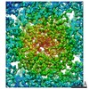
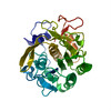
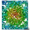
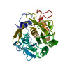
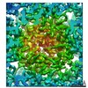
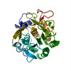
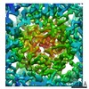
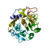
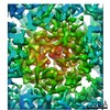
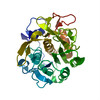
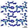
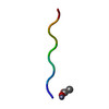
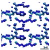
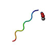
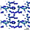
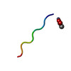
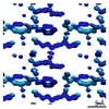
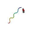
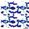
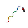
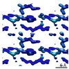
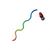
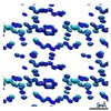
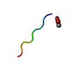
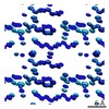
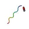
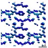
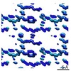
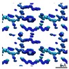
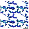
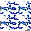
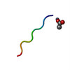
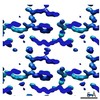
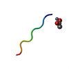
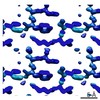
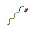
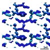
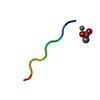
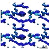
| タイトル | Analysis of Global and Site-Specific Radiation Damage in Cryo-EM. |
|---|---|
| ジャーナル・号・ページ | Structure, Vol. 26, Issue 5, Page 759-766.e4, Year 2018 |
| 掲載日 | 2018年5月1日 |
 著者 著者 | Johan Hattne / Dan Shi / Calina Glynn / Chih-Te Zee / Marcus Gallagher-Jones / Michael W Martynowycz / Jose A Rodriguez / Tamir Gonen /  |
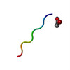
| PubMed 要旨 | Micro-crystal electron diffraction (MicroED) combines the efficiency of electron scattering with diffraction to allow structure determination from nano-sized crystalline samples in cryoelectron ...Micro-crystal electron diffraction (MicroED) combines the efficiency of electron scattering with diffraction to allow structure determination from nano-sized crystalline samples in cryoelectron microscopy (cryo-EM). It has been used to solve structures of a diverse set of biomolecules and materials, in some cases to sub-atomic resolution. However, little is known about the damaging effects of the electron beam on samples during such measurements. We assess global and site-specific damage from electron radiation on nanocrystals of proteinase K and of a prion hepta-peptide and find that the dynamics of electron-induced damage follow well-established trends observed in X-ray crystallography. Metal ions are perturbed, disulfide bonds are broken, and acidic side chains are decarboxylated while the diffracted intensities decay exponentially with increasing exposure. A better understanding of radiation damage in MicroED improves our assessment and processing of all types of cryo-EM data. |
 リンク リンク | Structure / PubMed:29706530 / PubMed Central |
| 手法 | EM (電子線結晶学) |
| 解像度 | 1.01 - 3.2 Å |
| 構造データ | EMDB-7490, PDB-6cl7: EMDB-7491, PDB-6cl8: EMDB-7492, PDB-6cl9: EMDB-7493, PDB-6cla: EMDB-7494, PDB-6clb: EMDB-7495, PDB-6clc: EMDB-7496, PDB-6cld: EMDB-7497, PDB-6cle: EMDB-7498, PDB-6clf: EMDB-7499, PDB-6clg: EMDB-7500, PDB-6clh: EMDB-7501, PDB-6cli: EMDB-7502, PDB-6clj: EMDB-7503, PDB-6clk: EMDB-7504, PDB-6cll: EMDB-7505, PDB-6clm: EMDB-7506, PDB-6cln: EMDB-7507, PDB-6clo: EMDB-7508, PDB-6clp: EMDB-7509, PDB-6clq: EMDB-7510, PDB-6clr: |
| 化合物 |  ChemComp-ACT:  ChemComp-ZN:  ChemComp-HOH: |
| 由来 |
|
 キーワード キーワード | HYDROLASE / PROTEIN FIBRIL / Amyloid fibril / prion / zinc binding |


 Engyodontium album (菌類)