 Movie
Movie Controller


+ Open data

- Basic information
| Entry | Database: PDB / ID: 6clk | ||||||
|---|---|---|---|---|---|---|---|
| Title | 1.01 A MicroED structure of GSNQNNF at 0.82 e- / A^2 | ||||||
 Components Components | GSNQNNF | ||||||
 Keywords Keywords | PROTEIN FIBRIL / Amyloid fibril / prion / zinc binding | ||||||
| Function / homology | ACETATE ION Function and homology information Function and homology information | ||||||
| Biological species | synthetic construct (others) | ||||||
| Method | ELECTRON CRYSTALLOGRAPHY / electron crystallography / cryo EM / Resolution: 1.01 Å | ||||||
 Authors Authors | Hattne, J. / Shi, D. / Glynn, C. / Zee, C.-T. / Gallagher-Jones, M. / Martynowycz, M.W. / Rodriguez, J.A. / Gonen, T. | ||||||
 Citation Citation | Journal: Structure / Year: 2018 Title: Analysis of Global and Site-Specific Radiation Damage in Cryo-EM. Authors: Johan Hattne / Dan Shi / Calina Glynn / Chih-Te Zee / Marcus Gallagher-Jones / Michael W Martynowycz / Jose A Rodriguez / Tamir Gonen /  Abstract: Micro-crystal electron diffraction (MicroED) combines the efficiency of electron scattering with diffraction to allow structure determination from nano-sized crystalline samples in cryoelectron ...Micro-crystal electron diffraction (MicroED) combines the efficiency of electron scattering with diffraction to allow structure determination from nano-sized crystalline samples in cryoelectron microscopy (cryo-EM). It has been used to solve structures of a diverse set of biomolecules and materials, in some cases to sub-atomic resolution. However, little is known about the damaging effects of the electron beam on samples during such measurements. We assess global and site-specific damage from electron radiation on nanocrystals of proteinase K and of a prion hepta-peptide and find that the dynamics of electron-induced damage follow well-established trends observed in X-ray crystallography. Metal ions are perturbed, disulfide bonds are broken, and acidic side chains are decarboxylated while the diffracted intensities decay exponentially with increasing exposure. A better understanding of radiation damage in MicroED improves our assessment and processing of all types of cryo-EM data. | ||||||
| History |
|
- Structure visualization
| Movie |
Movie viewer |
|---|---|
| Structure viewer | Molecule: MolmilJmol/JSmol |
- Downloads & links
-Download
| PDBx/mmCIF format | 6clk.cif.gz | 11.7 KB | Display | PDBx/mmCIF format |
|---|---|---|---|---|
| PDB format | pdb6clk.ent.gz | 5.3 KB | Display | PDB format |
| PDBx/mmJSON format | 6clk.json.gz | Tree view | PDBx/mmJSON format | |
| Others |  Other downloads Other downloads |
-Validation report
| Arichive directory | https://data.pdbj.org/pub/pdb/validation_reports/cl/6clkftp://data.pdbj.org/pub/pdb/validation_reports/cl/6clk | HTTPS FTP |
|---|
-Related structure data
| Related structure data | 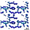 7503MC 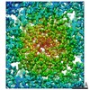 7490C 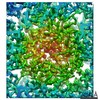 7491C 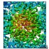 7492C 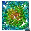 7493C 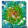 7494C 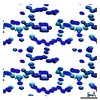 7495C 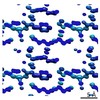 7496C  7497C  7498C  7499C  7500C  7501C  7502C  7504C  7505C  7506C  7507C  7508C  7509C 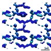 7510C 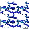 7511C 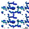 7512C 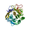 6cl7C  6cl8C  6cl9C  6claC  6clbC  6clcC  6cldC  6cleC  6clfC  6clgC  6clhC  6cliC  6cljC  6cllC  6clmC  6clnC  6cloC  6clpC  6clqC 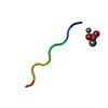 6clrC 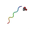 6clsC  6cltC M: map data used to model this data C: citing same article ( |
|---|---|
| Similar structure data |
-Links
 PDBj
PDBj

- Assembly
| Deposited unit | 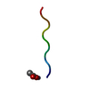
| ||||||||
|---|---|---|---|---|---|---|---|---|---|
| 1 |
| ||||||||
| Unit cell |
|
-Components
| #1: Protein/peptide | Mass: 779.756 Da / Num. of mol.: 1 / Source method: obtained synthetically / Source: (synth.) synthetic construct (others) |
|---|---|
| #2: Chemical | ChemComp-ACT /   Mass: 59.044 Da / Num. of mol.: 1 / Source method: obtained synthetically / Formula: C2H3O2 Mass: 59.044 Da / Num. of mol.: 1 / Source method: obtained synthetically / Formula: C2H3O2 |
| #3: Chemical | ChemComp-ZN /   Mass: 65.409 Da / Num. of mol.: 1 / Source method: obtained synthetically / Formula: Zn Mass: 65.409 Da / Num. of mol.: 1 / Source method: obtained synthetically / Formula: Zn |
| #4: Water | ChemComp-HOH /  Mass: 18.015 Da / Num. of mol.: 3 / Source method: isolated from a natural source / Formula: H2O Mass: 18.015 Da / Num. of mol.: 3 / Source method: isolated from a natural source / Formula: H2O |
-Experimental details
-Experiment
| Experiment | Method: ELECTRON CRYSTALLOGRAPHY |
|---|---|
| EM experiment | Aggregation state: 3D ARRAY / 3D reconstruction method: electron crystallography |
- Sample preparation
| Component | Name: Synthetic proto-filament / Type: COMPLEX / Entity ID: #1 / Source: MULTIPLE SOURCES | |||||||||||||||
|---|---|---|---|---|---|---|---|---|---|---|---|---|---|---|---|---|
| Molecular weight | Value: 0.000899141 MDa / Experimental value: NO | |||||||||||||||
| Buffer solution | pH: 6 | |||||||||||||||
| Buffer component |
| |||||||||||||||
| Specimen | Conc.: 10 mg/ml / Embedding applied: NO / Shadowing applied: NO / Staining applied: NO / Vitrification applied: YES | |||||||||||||||
| Specimen support | Grid material: COPPER / Grid mesh size: 300 divisions/in. / Grid type: Quantifoil R2/2 | |||||||||||||||
| Vitrification | Instrument: FEI VITROBOT MARK IV / Cryogen name: ETHANE / Humidity: 30 % |
-Data collection
| Experimental equipment |  Model: Tecnai F20 / Image courtesy: FEI Company |
|---|---|
| Microscopy | Model: FEI TECNAI F20 |
| Electron gun | Electron source:  FIELD EMISSION GUN / Accelerating voltage: 200 kV / Illumination mode: FLOOD BEAM FIELD EMISSION GUN / Accelerating voltage: 200 kV / Illumination mode: FLOOD BEAM |
| Electron lens | Mode: DIFFRACTION |
| Specimen holder | Cryogen: NITROGEN Specimen holder model: GATAN 626 SINGLE TILT LIQUID NITROGEN CRYO TRANSFER HOLDER |
| Image recording | Average exposure time: 2.1 sec. / Electron dose: 0.00357 e/Å2 / Film or detector model: TVIPS TEMCAM-F416 (4k x 4k) / Num. of diffraction images: 826 / Num. of grids imaged: 1 / Num. of real images: 826 |
| Image scans | Sampling size: 31.2 µm / Width: 2048 / Height: 2048 |
| EM diffraction | Camera length: 730 mm |
| EM diffraction shell | Resolution: 1.01→1.04 Å / Fourier space coverage: 77.99 % / Multiplicity: 4.3 / Num. of structure factors: 124 / Phase residual: 62.42 ° |
| EM diffraction stats | Fourier space coverage: 80 % / High resolution: 1.01 Å / Num. of intensities measured: 12579 / Num. of structure factors: 1952 / Phase error: 42.71 ° / Phase residual: 42.71 ° / Phase error rejection criteria: 0 / Rmerge: 0.289 / Rsym: 0.289 |
| Detector | Date: Aug 1, 2017 |
- Processing
| Software |
| ||||||||||||||||||||||||||||||||||||||||||||||||||||||||||||||||||||||||||||||||||||||||||||||||||||||||||
|---|---|---|---|---|---|---|---|---|---|---|---|---|---|---|---|---|---|---|---|---|---|---|---|---|---|---|---|---|---|---|---|---|---|---|---|---|---|---|---|---|---|---|---|---|---|---|---|---|---|---|---|---|---|---|---|---|---|---|---|---|---|---|---|---|---|---|---|---|---|---|---|---|---|---|---|---|---|---|---|---|---|---|---|---|---|---|---|---|---|---|---|---|---|---|---|---|---|---|---|---|---|---|---|---|---|---|---|
| EM software |
| ||||||||||||||||||||||||||||||||||||||||||||||||||||||||||||||||||||||||||||||||||||||||||||||||||||||||||
| EM 3D crystal entity | ∠α: 83.65 ° / ∠β: 85.53 ° / ∠γ: 82.814 ° / A: 4.84 Å / B: 14.1 Å / C: 17.38 Å / Space group name: P1 / Space group num: 1 | ||||||||||||||||||||||||||||||||||||||||||||||||||||||||||||||||||||||||||||||||||||||||||||||||||||||||||
| CTF correction | Type: NONE | ||||||||||||||||||||||||||||||||||||||||||||||||||||||||||||||||||||||||||||||||||||||||||||||||||||||||||
| 3D reconstruction | Resolution: 1.01 Å / Resolution method: DIFFRACTION PATTERN/LAYERLINES / Symmetry type: 3D CRYSTAL | ||||||||||||||||||||||||||||||||||||||||||||||||||||||||||||||||||||||||||||||||||||||||||||||||||||||||||
| Atomic model building | B value: 4.322 / Protocol: OTHER / Space: RECIPROCAL / Details: Electron scattering factors | ||||||||||||||||||||||||||||||||||||||||||||||||||||||||||||||||||||||||||||||||||||||||||||||||||||||||||
| Refinement | Resolution: 1.01→13.92 Å / Cor.coef. Fo:Fc: 0.887 / Cor.coef. Fo:Fc free: 0.884 / SU B: 1.01 / SU ML: 0.052 / Cross valid method: THROUGHOUT / ESU R: 0.061 / ESU R Free: 0.057 / Details: HYDROGENS HAVE BEEN ADDED IN THE RIDING POSITIONS
| ||||||||||||||||||||||||||||||||||||||||||||||||||||||||||||||||||||||||||||||||||||||||||||||||||||||||||
| Solvent computation | Ion probe radii: 0.8 Å / Shrinkage radii: 0.8 Å / VDW probe radii: 1.2 Å | ||||||||||||||||||||||||||||||||||||||||||||||||||||||||||||||||||||||||||||||||||||||||||||||||||||||||||
| Displacement parameters | Biso mean: 4.322 Å2
| ||||||||||||||||||||||||||||||||||||||||||||||||||||||||||||||||||||||||||||||||||||||||||||||||||||||||||
| Refinement step | Cycle: 1 / Total: 63 | ||||||||||||||||||||||||||||||||||||||||||||||||||||||||||||||||||||||||||||||||||||||||||||||||||||||||||
| Refine LS restraints |
|