Movie
Movie Controller
 Structure viewers
 About Yorodumi Papers


+Search query
-Structure paper
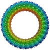
| Title | Cryo-EM structure of the gasdermin A3 membrane pore. |
|---|---|
| Journal, issue, pages | Nature, Vol. 557, Issue 7703, Page 62-67, Year 2018 |
| Publish date | Apr 25, 2018 |
 Authors Authors | Jianbin Ruan / Shiyu Xia / Xing Liu / Judy Lieberman / Hao Wu /  |
| PubMed Abstract | Gasdermins mediate inflammatory cell death after cleavage by caspases or other, unknown enzymes. The cleaved N-terminal fragments bind to acidic membrane lipids to form pores, but the mechanism of ...Gasdermins mediate inflammatory cell death after cleavage by caspases or other, unknown enzymes. The cleaved N-terminal fragments bind to acidic membrane lipids to form pores, but the mechanism of pore formation remains unresolved. Here we present the cryo-electron microscopy structures of the 27-fold and 28-fold single-ring pores formed by the N-terminal fragment of mouse GSDMA3 (GSDMA3-NT) at 3.8 and 4.2 Å resolutions, and of a double-ring pore at 4.6 Å resolution. In the 27-fold pore, a 108-stranded anti-parallel β-barrel is formed by two β-hairpins from each subunit capped by a globular domain. We identify a positively charged helix that interacts with the acidic lipid cardiolipin. GSDMA3-NT undergoes radical conformational changes upon membrane insertion to form long, membrane-spanning β-strands. We also observe an unexpected additional symmetric ring of GSDMA3-NT subunits that does not insert into the membrane in the double-ring pore, which may represent a pre-pore state of GSDMA3-NT. These structures provide a basis that explains the activities of several mutant gasdermins, including defective mutants that are associated with cancer. |
 External links External links | Nature / PubMed:29695864 / PubMed Central |
| Methods | EM (single particle) |
| Resolution | 3.8 - 4.6 Å |
| Structure data | EMDB-7449, PDB-6cb8: 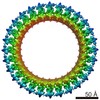 EMDB-7450: 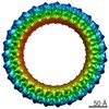 EMDB-7451: |
| Chemicals |  ChemComp-CDL: |
| Source |
|
 Keywords Keywords | IMMUNE SYSTEM / Pyroptosis / Pore forming protein |