[English] 日本語
Yorodumi Papers
- Database of articles cited by EMDB/PDB/SASBDB data -

+
Search query

Keywords
Structure methods
Author
Journal
IF

-
Structure paper

TitleNucleosome spacing regulates linker methylation by DNMT3A2/3B3.
Journal, issue, pagesMol Cell, Vol. 86, Issue 5, Page 834-850.e9, Year 2026
Publish dateMar 5, 2026
AuthorsXiaoyan Xie / Minmin Liu / Gabriella N L Chua / X Edward Zhou / Michelle L Dykstra / Shixin Liu / Peter A Jones / Evan J Worden /
PubMed AbstractDe novo CpG methylation (mCpG) is deposited by DNMT3A and DNMT3B, which target DNA linkers between nucleosomes. Cells contain millions of unique linkers, but the rules dictating which linkers get ...De novo CpG methylation (mCpG) is deposited by DNMT3A and DNMT3B, which target DNA linkers between nucleosomes. Cells contain millions of unique linkers, but the rules dictating which linkers get targeted by DNMT3 enzymes are not understood. We show that nucleosome spacing controls linker DNA methylation and H3K36me2 recognition by human DNMT3A2/3B3, linking de novo methylation to chromatin architecture. We present structures of DNMT3A2/3B3 bound to dinucleosomes, revealing that short linkers promote dinucleosome bridging, blocking access to linker DNA and suppressing methylation, whereas long linkers allow DNMT3A2/3B3 to engage each nucleosome separately and methylate linker DNA. Finally, we show that DNMT3A2/3B3 positions proline-tryptophan-tryptophan-proline (PWWP) domains to scan for H3K36me2. However, H3K36me2 recognition is blocked when DNMT3A2/3B3 bridges dinucleosomes with short linkers, imposing an additional structural constraint on DNMT3A2/3B3 function. Together, these findings uncover the mechanisms that govern de novo methylation in chromatin and explain how DNMT3 enzymes target linkers in cells.
External linksMol Cell / PubMed:41742418 / PubMed Central
MethodsEM (single particle)
Resolution3.14 - 8.0 Å
Structure data

EMDB-47344, PDB-9e00:
Cryo-EM structure of human DNMT3A2-DNMT3B3 complex bound to di-nucleosome
Method: EM (single particle) / Resolution: 3.75 Å

EMDB-47349, PDB-9e05:
The consensus model of the cryo-EM structure of human DNMT3A2-DNMT3B3 complex bound to di-nucleosome
Method: EM (single particle) / Resolution: 8.0 Å

EMDB-47353, PDB-9e09:
Cryo-EM structure a single nucleosome (2) focus of human DNMT3A2-DNMT3B3 complex bound to di-nucleosome
Method: EM (single particle) / Resolution: 3.72 Å

EMDB-47354, PDB-9e0f:
A focus of DNMT tetramer (1) of the cryo-EM structure of human DNMT3A2-DNMT3B3 complex bound to di-nucleosome
Method: EM (single particle) / Resolution: 3.46 Å

EMDB-47355, PDB-9e0g:
A focus of tetramer (2) of the cryo-EM structure of human DNMT3A2-DNMT3B3 complex bound to di-nucleosome
Method: EM (single particle) / Resolution: 3.72 Å

EMDB-47448, PDB-9e2d:
Cryo-EM structure of human DNMT3A2-DNMT3B3 complex bound to a di-nucleosome with a five base pair linker
Method: EM (single particle) / Resolution: 6.08 Å

EMDB-47450, PDB-9e2f:
Cryo-EM structure of a di-nucleosome with a five base pair linker
Method: EM (single particle) / Resolution: 5.9 Å

EMDB-47461, PDB-9e2q:
Cryo-EM structure of DNMT 3A2/3B3 tetramer in complex with a di-nucleosome with a six base pair linker
Method: EM (single particle) / Resolution: 3.14 Å

EMDB-47462, PDB-9e2r:
Cryo-EM structure of human DNMT3A2-DNMT3B3 complex bound to a di-nucleosome and an histone-3 peptide
Method: EM (single particle) / Resolution: 7.71 Å

EMDB-47479, PDB-9e3d:
Cryo-EM structure of DNMT 3A2/3B3 tetramer in complex with a di-nucleosome with K120R mutant H2B
Method: EM (single particle) / Resolution: 3.8 Å

EMDB-47495, PDB-9e3r:
Cryo-EM structure of PWWP domain deleted DNMT 3A2/3B3 in complex with a di-nucleosome
Method: EM (single particle) / Resolution: 6.9 Å

EMDB-47499, PDB-9e49:
Cryo-EM structure of a di-nucleosome with an eight base-pair linker
Method: EM (single particle) / Resolution: 5.93 Å

EMDB-71301: Consensus map for Cryo-EM structure of DNMT3A2/3B3 tetramer in complex with a 167H3K36me2 nucleosome
Method: EM (single particle) / Resolution: 3.63 Å

EMDB-71302: Focus map of DNMT3A2/3B3 tetramer for Cryo-EM structure of DNMT3A2/3B3 in complex with 167H3K36me2-nucleosome
Method: EM (single particle) / Resolution: 6.35 Å

EMDB-71304: Focus map of PWWP_1 domain for Cryo-EM structure of DNMT3A2/3B3 in complex with 167H3K36me2-nucleosome
Method: EM (single particle) / Resolution: 4.91 Å

EMDB-71305: Focus map of PWWP_2 for Cryo-EM structure of DNMT3A2/3B3 in complex with 167H3K36me2-nucleosome
Method: EM (single particle) / Resolution: 5.86 Å

EMDB-71306: Focus map of nucleosome for Cryo-EM structure of DNMT3A2/3B3 in complex with 167H3K36me2-nucleosome
Method: EM (single particle) / Resolution: 4.43 Å

EMDB-71604: Consensus map for Cryo-EM structure of DNMT3A2/3B3 in complex with H3K36me2 di-nucleosome with eight-base-pair linker
Method: EM (single particle) / Resolution: 3.84 Å

EMDB-71605: Focus map of DNMT3A2/3B3 tetramer1 for Cryo-EM structure of DNMT3A2/3B3 in complex with H3K36me2 di-nucleosome with eight base pair linker
Method: EM (single particle) / Resolution: 4.89 Å

EMDB-71606: Focus map of DNMT3A2/3B3 tetramer2 for Cryo-EM structure of DNMT3A2/3B3 in complex with H3K36me2 di-nucleosome with eight base pair linker
Method: EM (single particle) / Resolution: 5.51 Å

EMDB-71607: Focus map of nucleosome 1 for Cryo-EM structure of DNMT3A2/3B3 in complex with H3K36me2 di-nucleosome with eight base pair linker
Method: EM (single particle) / Resolution: 4.99 Å

EMDB-71608: Focus map of nucleosome 2 for Cryo-EM structure of DNMT3A2/3B3 in complex with H3K36me2 di-nucleosome with eight base pair linker
Method: EM (single particle) / Resolution: 3.51 Å

EMDB-71609: Focus map of nucleosome1_PWWP1 for Cryo-EM structure of DNMT3A2/3B3 in complex with H3K36me2 di-nucleosome with eight base pair linker
Method: EM (single particle) / Resolution: 4.99 Å

EMDB-72318, PDB-9q7u:
Composite map for Cryo-EM structure of DNMT3A2-DNMT3B3 tetramer bound to 167H3K36me2-nucleosome
Method: EM (single particle) / Resolution: 3.4 Å

EMDB-72487, PDB-9y4p:
Cryo-EM structure of DNMT3A2/3B3 in complex with H3K36me2 di-nucleosome with eight base pair linker
Method: EM (single particle) / Resolution: 3.84 Å

Chemicals

ChemComp-ZN:
Unknown entry

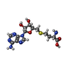
ChemComp-SAH:
S-ADENOSYL-L-HOMOCYSTEINE

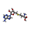
ChemComp-SAO:
5'-S-[(3S)-3-azaniumyl-3-carboxypropyl]-5'-thioadenosine

Source
  • homo sapiens (human)
  • xenopus laevis (African clawed frog)
KeywordsDNA BINDING PROTEIN / DNMT3A2 / DNMT3B3 / DNA methylation / di-nucleosome / DNA BINDING PROTEIN/DNA / DNMT3A / DNMT3B / nucleosome / DNA BINDING PROTEIN-DNA complex / DNMT 3A2/3B3 / methylation / DNMT3A2-DNMT3B3 / 167H3K36me2-nucleosome / DNMT3A2/DNMT3B3 / H3K36me2 di-nucleosome

+
About Yorodumi Papers

-
News

-
Feb 9, 2022. New format data for meta-information of EMDB entries

New format data for meta-information of EMDB entries

  • Version 3 of the EMDB header file is now the official format.
  • The previous official version 1.9 will be removed from the archive.

Related info.:EMDB header

External links:wwPDB to switch to version 3 of the EMDB data model

-
Aug 12, 2020. Covid-19 info

Covid-19 info

URL: https://pdbj.org/emnavi/covid19.php

New page: Covid-19 featured information page in EM Navigator.

Related info.:Covid-19 info / Mar 5, 2020. Novel coronavirus structure data

+
Mar 5, 2020. Novel coronavirus structure data

Novel coronavirus structure data

Related info.:Yorodumi Speices / Aug 12, 2020. Covid-19 info

External links:COVID-19 featured content - PDBj / Molecule of the Month (242):Coronavirus Proteases

+
Jan 31, 2019. EMDB accession codes are about to change! (news from PDBe EMDB page)

EMDB accession codes are about to change! (news from PDBe EMDB page)

  • The allocation of 4 digits for EMDB accession codes will soon come to an end. Whilst these codes will remain in use, new EMDB accession codes will include an additional digit and will expand incrementally as the available range of codes is exhausted. The current 4-digit format prefixed with “EMD-” (i.e. EMD-XXXX) will advance to a 5-digit format (i.e. EMD-XXXXX), and so on. It is currently estimated that the 4-digit codes will be depleted around Spring 2019, at which point the 5-digit format will come into force.
  • The EM Navigator/Yorodumi systems omit the EMD- prefix.

Related info.:Q: What is EMD? / ID/Accession-code notation in Yorodumi/EM Navigator

External links:EMDB Accession Codes are Changing Soon! / Contact to PDBj

+
Jul 12, 2017. Major update of PDB

Major update of PDB

  • wwPDB released updated PDB data conforming to the new PDBx/mmCIF dictionary.
  • This is a major update changing the version number from 4 to 5, and with Remediation, in which all the entries are updated.
  • In this update, many items about electron microscopy experimental information are reorganized (e.g. em_software).
  • Now, EM Navigator and Yorodumi are based on the updated data.

External links:wwPDB Remediation / Enriched Model Files Conforming to OneDep Data Standards Now Available in the PDB FTP Archive

-
Yorodumi Papers

Database of articles cited by EMDB/PDB/SASBDB data

  • Database of articles cited by EMDB, PDB, and SASBDB entries
  • Using PubMed data

Related info.:EMDB / PDB / SASBDB / Yorodumi / EMN Papers / Changes in new EM Navigator and Yorodumi

Read more