[English] 日本語
Yorodumi Papers
- Database of articles cited by EMDB/PDB/SASBDB data -

+
Search query

Keywords
Structure methods
Author
Journal
IF

-
Structure paper

TitleStructural mechanism of 3'3'-cGAMP-induced filamentation and phospholipid hydrolysis by CapV in bacterial antiphage defense.
Journal, issue, pagesCell Rep, Vol. 45, Issue 4, Page 117261, Year 2026
Publish dateApr 13, 2026
AuthorsYun Lv / Sheng Liu / Qihai Wang / Jing Zhu / Yingxiang Hou / Haiyan Xu / Deyan Zhu / Yu Liu / Jing Wu / Changxin Wu / Guijun Shang / Hongxiang Lou / Defen Lu / Huiqing Yuan / Deyu Zhu /
PubMed AbstractThe cyclic-oligonucleotide-based antiphage signaling system (CBASS) protects bacteria from phage infection. In Vibrio cholerae, phage infection activates CD-NTase DncV to produce 3'3'-cGAMP, which ...The cyclic-oligonucleotide-based antiphage signaling system (CBASS) protects bacteria from phage infection. In Vibrio cholerae, phage infection activates CD-NTase DncV to produce 3'3'-cGAMP, which triggers phospholipase CapV to degrade phosphatidylethanolamine and phosphatidylglycerol, the major phospholipids in the inner-membranes, thereby inducing cell death. However, how 3'3'-cGAMP activates CapV was unclear. Here we present crystal structures of inactive Acinetobacter baumannii CapV in apo and 3'3'-cGAMP-bound forms, along with cryo-EM structures of activated CapV-3'3'-cGAMP complex, with or without substrate dioleoylphosphatidyl-ethanolamine (DOPE). Apo-CapV forms symmetric dimers in a "closed" state. 3'3'-cGAMP binding drives lateral polymerization of dimers into filament assembly, inducing an "open" state that exposes the active site and substrate-binding cleft. DOPE binding further shifts CapV to an "ajar" state, where a Y-shaped cleft positions DOPE for hydrolysis via a conserved Ser/Asp catalytic dyad. This 3'3'-cGAMP-induced filamentation mirrors activation mechanisms of TIR-STING, TIR-SAVED, and mammalian STING, revealing a conserved signaling pattern across immune systems.
External linksCell Rep / PubMed:41984589
MethodsEM (single particle) / X-ray diffraction
Resolution2 - 3.0 Å
Structure data

EMDB-62333, PDB-9kh6:
cryo-EM structure of lipase/ligand/substrate complex
Method: EM (single particle) / Resolution: 2.8 Å

EMDB-62334: Cryo-EM structure of lipase/ligand complex
PDB-9kh7: cryo-EM structure of lipase/ligand complex
Method: EM (single particle) / Resolution: 3.0 Å

PDB-9wn0:
Crystal structure of phospholipase A2
Method: X-RAY DIFFRACTION / Resolution: 2 Å

PDB-9wn1:
PhospholipaseA2 with a ligand
Method: X-RAY DIFFRACTION / Resolution: 2.76 Å

Chemicals

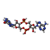
ChemComp-4BW:
2-amino-9-[(2R,3R,3aS,5R,7aR,9R,10R,10aS,12R,14aR)-9-(6-amino-9H-purin-9-yl)-3,5,10,12-tetrahydroxy-5,12-dioxidooctahydro-2H,7H-difuro[3,2-d:3',2'-j][1,3,7,9,2,8]tetraoxadiphosphacyclododecin-2-yl]-1,9-dihydro-6H-purin-6-one

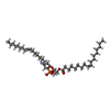
ChemComp-PEE:
1,2-dioleoyl-sn-glycero-3-phosphoethanolamine / DOPE, phospholipid*YM

ChemComp-HOH:
WATER

Source
  • acinetobacter baumannii (bacteria)
KeywordsHYDROLASE / lipase / LIPID BINDING PROTEIN / phospholipase / crystal sturcure / phospholipid / phospholipasec2 / ligand

+
About Yorodumi Papers

-
News

-
Feb 9, 2022. New format data for meta-information of EMDB entries

New format data for meta-information of EMDB entries

  • Version 3 of the EMDB header file is now the official format.
  • The previous official version 1.9 will be removed from the archive.

Related info.:EMDB header

External links:wwPDB to switch to version 3 of the EMDB data model

-
Aug 12, 2020. Covid-19 info

Covid-19 info

URL: https://pdbj.org/emnavi/covid19.php

New page: Covid-19 featured information page in EM Navigator.

Related info.:Covid-19 info / Mar 5, 2020. Novel coronavirus structure data

+
Mar 5, 2020. Novel coronavirus structure data

Novel coronavirus structure data

Related info.:Yorodumi Speices / Aug 12, 2020. Covid-19 info

External links:COVID-19 featured content - PDBj / Molecule of the Month (242):Coronavirus Proteases

+
Jan 31, 2019. EMDB accession codes are about to change! (news from PDBe EMDB page)

EMDB accession codes are about to change! (news from PDBe EMDB page)

  • The allocation of 4 digits for EMDB accession codes will soon come to an end. Whilst these codes will remain in use, new EMDB accession codes will include an additional digit and will expand incrementally as the available range of codes is exhausted. The current 4-digit format prefixed with “EMD-” (i.e. EMD-XXXX) will advance to a 5-digit format (i.e. EMD-XXXXX), and so on. It is currently estimated that the 4-digit codes will be depleted around Spring 2019, at which point the 5-digit format will come into force.
  • The EM Navigator/Yorodumi systems omit the EMD- prefix.

Related info.:Q: What is EMD? / ID/Accession-code notation in Yorodumi/EM Navigator

External links:EMDB Accession Codes are Changing Soon! / Contact to PDBj

+
Jul 12, 2017. Major update of PDB

Major update of PDB

  • wwPDB released updated PDB data conforming to the new PDBx/mmCIF dictionary.
  • This is a major update changing the version number from 4 to 5, and with Remediation, in which all the entries are updated.
  • In this update, many items about electron microscopy experimental information are reorganized (e.g. em_software).
  • Now, EM Navigator and Yorodumi are based on the updated data.

External links:wwPDB Remediation / Enriched Model Files Conforming to OneDep Data Standards Now Available in the PDB FTP Archive

-
Yorodumi Papers

Database of articles cited by EMDB/PDB/SASBDB data

  • Database of articles cited by EMDB, PDB, and SASBDB entries
  • Using PubMed data

Related info.:EMDB / PDB / SASBDB / Yorodumi / EMN Papers / Changes in new EM Navigator and Yorodumi

Read more