 Movie
Movie Controller


+ Open data

- Basic information
| Entry | Database: PDB / ID: 9wn1 | ||||||
|---|---|---|---|---|---|---|---|
| Title | PhospholipaseA2 with a ligand | ||||||
 Components Components | Patatin | ||||||
 Keywords Keywords | LIPID BINDING PROTEIN / phospholipasec2 / crystal sturcure / ligand | ||||||
| Function / homology | : / Patatin-like phospholipase domain / Patatin-like phospholipase / Patatin-like phospholipase (PNPLA) domain profile. / Acyl transferase/acyl hydrolase/lysophospholipase / lipid catabolic process / hydrolase activity / Chem-4BW / Patatin Function and homology information Function and homology information | ||||||
| Biological species |  Acinetobacter baumannii (bacteria) Acinetobacter baumannii (bacteria) | ||||||
| Method |  X-RAY DIFFRACTION / SYNCHROTRON / MOLECULAR REPLACEMENT / Resolution: 2.76 Å X-RAY DIFFRACTION / SYNCHROTRON / MOLECULAR REPLACEMENT / Resolution: 2.76 Å | ||||||
 Authors Authors | Zhu, D. / Wang, Q. | ||||||
| Funding support | 1items
| ||||||
 Citation Citation | Journal: Cell Rep / Year: 2026 Title: Structural mechanism of 3'3'-cGAMP-induced filamentation and phospholipid hydrolysis by CapV in bacterial antiphage defense. Authors: Yun Lv / Sheng Liu / Qihai Wang / Jing Zhu / Yingxiang Hou / Haiyan Xu / Deyan Zhu / Yu Liu / Jing Wu / Changxin Wu / Guijun Shang / Hongxiang Lou / Defen Lu / Huiqing Yuan / Deyu Zhu /  Abstract: The cyclic-oligonucleotide-based antiphage signaling system (CBASS) protects bacteria from phage infection. In Vibrio cholerae, phage infection activates CD-NTase DncV to produce 3'3'-cGAMP, which ...The cyclic-oligonucleotide-based antiphage signaling system (CBASS) protects bacteria from phage infection. In Vibrio cholerae, phage infection activates CD-NTase DncV to produce 3'3'-cGAMP, which triggers phospholipase CapV to degrade phosphatidylethanolamine and phosphatidylglycerol, the major phospholipids in the inner-membranes, thereby inducing cell death. However, how 3'3'-cGAMP activates CapV was unclear. Here we present crystal structures of inactive Acinetobacter baumannii CapV in apo and 3'3'-cGAMP-bound forms, along with cryo-EM structures of activated CapV-3'3'-cGAMP complex, with or without substrate dioleoylphosphatidyl-ethanolamine (DOPE). Apo-CapV forms symmetric dimers in a "closed" state. 3'3'-cGAMP binding drives lateral polymerization of dimers into filament assembly, inducing an "open" state that exposes the active site and substrate-binding cleft. DOPE binding further shifts CapV to an "ajar" state, where a Y-shaped cleft positions DOPE for hydrolysis via a conserved Ser/Asp catalytic dyad. This 3'3'-cGAMP-induced filamentation mirrors activation mechanisms of TIR-STING, TIR-SAVED, and mammalian STING, revealing a conserved signaling pattern across immune systems. | ||||||
| History |
|
- Structure visualization
| Structure viewer | Molecule: MolmilJmol/JSmol |
|---|
- Downloads & links
-Download
| PDBx/mmCIF format | 9wn1.cif.gz | 760.1 KB | Display | PDBx/mmCIF format |
|---|---|---|---|---|
| PDB format | pdb9wn1.ent.gz | 631.7 KB | Display | PDB format |
| PDBx/mmJSON format | 9wn1.json.gz | Tree view | PDBx/mmJSON format | |
| Others |  Other downloads Other downloads |
-Validation report
| Arichive directory | https://data.pdbj.org/pub/pdb/validation_reports/wn/9wn1ftp://data.pdbj.org/pub/pdb/validation_reports/wn/9wn1 | HTTPS FTP |
|---|
-Related structure data


-Links
 PDBj
PDBj
- Assembly
| Deposited unit | 
| ||||||||
|---|---|---|---|---|---|---|---|---|---|
| 1 | 
| ||||||||
| 2 | 
| ||||||||
| 3 | 
| ||||||||
| 4 | 
| ||||||||
| 5 | 
| ||||||||
| 6 | 
| ||||||||
| Unit cell |
|
-Components
| #1: Protein | Mass: 36882.965 Da / Num. of mol.: 12 / Mutation: K304A Source method: isolated from a genetically manipulated source Details: Sequence reference for strain 'Acinetobacter baumannii' is not available in UniProt at the time of biocuration. Current sequence reference is from UniProt id A0A4Q7ABV3. Source: (gene. exp.) Acinetobacter baumannii (bacteria) / Gene: EXU28_18015 / Production host: #2: Chemical | ChemComp-4BW / 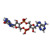  Mass: 674.411 Da / Num. of mol.: 12 / Source method: obtained synthetically / Formula: C20H24N10O13P2 / Feature type: SUBJECT OF INVESTIGATION Mass: 674.411 Da / Num. of mol.: 12 / Source method: obtained synthetically / Formula: C20H24N10O13P2 / Feature type: SUBJECT OF INVESTIGATION#3: Water | ChemComp-HOH / |  Mass: 18.015 Da / Num. of mol.: 196 / Source method: isolated from a natural source / Formula: H2O Mass: 18.015 Da / Num. of mol.: 196 / Source method: isolated from a natural source / Formula: H2OHas ligand of interest | Y | Has protein modification | N | |
|---|
-Experimental details
-Experiment
| Experiment | Method: X-RAY DIFFRACTION / Number of used crystals: 1 |
|---|
- Sample preparation
| Crystal | Density Matthews: 2.13 Å3/Da / Density % sol: 42.13 % |
|---|---|
| Crystal grow | Temperature: 293 K / Method: vapor diffusion, hanging drop / Details: 8% PEG6000,0.1M MES 6.0, 0.2M KCL |
-Data collection
| Diffraction | Mean temperature: 100 K / Serial crystal experiment: N |
|---|---|
| Diffraction source | Source: SYNCHROTRON / Site: SSRF / Beamline: BL19U1 / Wavelength: 0.9791 Å |
| Detector | Type: APEX II CCD / Detector: CCD / Date: Jan 25, 2023 |
| Radiation | Protocol: SINGLE WAVELENGTH / Monochromatic (M) / Laue (L): M / Scattering type: x-ray |
| Radiation wavelength | Wavelength: 0.9791 Å / Relative weight: 1 |
| Reflection | Resolution: 2.76→50 Å / Num. obs: 90466 / % possible obs: 95.5 % / Redundancy: 6.9 % / CC1/2: 0.994 / Net I/σ(I): 7.5 |
| Reflection shell | Resolution: 2.76→2.91 Å / Num. unique obs: 2802 / CC1/2: 0.774 |
- Processing
| Software |
| |||||||||||||||||||||||||||||||||||||||||||||||||||||||||||||||||||||||||||||||||||||||||||||||||||||||||||||||||||||||||||||||||||||||||||||||||||||||||||||||||||||||||||||||||||||||||||||||||||||||||||||||||||||||||
|---|---|---|---|---|---|---|---|---|---|---|---|---|---|---|---|---|---|---|---|---|---|---|---|---|---|---|---|---|---|---|---|---|---|---|---|---|---|---|---|---|---|---|---|---|---|---|---|---|---|---|---|---|---|---|---|---|---|---|---|---|---|---|---|---|---|---|---|---|---|---|---|---|---|---|---|---|---|---|---|---|---|---|---|---|---|---|---|---|---|---|---|---|---|---|---|---|---|---|---|---|---|---|---|---|---|---|---|---|---|---|---|---|---|---|---|---|---|---|---|---|---|---|---|---|---|---|---|---|---|---|---|---|---|---|---|---|---|---|---|---|---|---|---|---|---|---|---|---|---|---|---|---|---|---|---|---|---|---|---|---|---|---|---|---|---|---|---|---|---|---|---|---|---|---|---|---|---|---|---|---|---|---|---|---|---|---|---|---|---|---|---|---|---|---|---|---|---|---|---|---|---|---|---|---|---|---|---|---|---|---|---|---|---|---|---|---|---|---|
| Refinement | Method to determine structure: MOLECULAR REPLACEMENT / Resolution: 2.76→20.3 Å / SU ML: 0.38 / Cross valid method: FREE R-VALUE / σ(F): 1.34 / Phase error: 29.29 / Stereochemistry target values: ML
| |||||||||||||||||||||||||||||||||||||||||||||||||||||||||||||||||||||||||||||||||||||||||||||||||||||||||||||||||||||||||||||||||||||||||||||||||||||||||||||||||||||||||||||||||||||||||||||||||||||||||||||||||||||||||
| Solvent computation | Shrinkage radii: 0.9 Å / VDW probe radii: 1.11 Å / Solvent model: FLAT BULK SOLVENT MODEL | |||||||||||||||||||||||||||||||||||||||||||||||||||||||||||||||||||||||||||||||||||||||||||||||||||||||||||||||||||||||||||||||||||||||||||||||||||||||||||||||||||||||||||||||||||||||||||||||||||||||||||||||||||||||||
| Refinement step | Cycle: LAST / Resolution: 2.76→20.3 Å
| |||||||||||||||||||||||||||||||||||||||||||||||||||||||||||||||||||||||||||||||||||||||||||||||||||||||||||||||||||||||||||||||||||||||||||||||||||||||||||||||||||||||||||||||||||||||||||||||||||||||||||||||||||||||||
| Refine LS restraints |
| |||||||||||||||||||||||||||||||||||||||||||||||||||||||||||||||||||||||||||||||||||||||||||||||||||||||||||||||||||||||||||||||||||||||||||||||||||||||||||||||||||||||||||||||||||||||||||||||||||||||||||||||||||||||||
| LS refinement shell |
|
