 Movie
Movie Controller
 Structure viewers
 About Yorodumi Papers


+Search query
-Structure paper
| Title | Furin cleavage of the Moloney murine leukemia virus Env precursor reorganizes the spike structure. |
|---|---|
| Journal, issue, pages | Proc Natl Acad Sci U S A, Vol. 111, Issue 16, Page 6034-6039, Year 2014 |
| Publish date | Apr 22, 2014 |
 Authors Authors | Mathilda Sjöberg / Shang-Rung Wu / Robin Löving / Kimmo Rantalainen / Birgitta Lindqvist / Henrik Garoff /  |
| PubMed Abstract | The trimeric Moloney murine leukemia virus Env protein matures by two proteolytic cleavages. First, furin cleaves the Env precursor into the surface (SU) and transmembrane (TM) subunits in the cell ...The trimeric Moloney murine leukemia virus Env protein matures by two proteolytic cleavages. First, furin cleaves the Env precursor into the surface (SU) and transmembrane (TM) subunits in the cell and then the viral protease cleaves the R-peptide from TM in new virus. Here we analyzed the structure of the furin precursor, by cryoelectron microscopy. We transfected 293T cells with a furin cleavage site provirus mutant, R466G/K468G, and produced the virus in the presence of amprenavir to also inhibit the R-peptide cleavage. Although Env incorporation into particles was inhibited, enough precursor could be isolated and analyzed by cryoelectron microscopy to yield a 3D structure at 22 Å resolution. This showed an open cage-like structure like that of the R-peptide precursor and the mature Env described before. However, the middle protrusion of the protomeric unit, so prominently pointing out from the side of the more mature forms of the Env, was absent. Instead, there was extra density in the top protrusion. This suggested that the C-terminal SU domain was associated alongside the receptor binding N-terminal SU domain in the furin precursor. This was supported by mapping with a SU C-terminal domain-specific antigen binding fragment. We concluded that furin cleavage not only separates the subunits and liberates the fusion peptide at the end of TM but also allows the C-terminal domain to relocate into a peripheral position. This conformational change might explain how the C-terminal domain of SU gains the potential to undergo disulfide isomerization, an event that facilitates membrane fusion. |
 External links External links | Proc Natl Acad Sci U S A / PubMed:24711391 / PubMed Central |
| Methods | EM (single particle) |
| Resolution | 22.0 - 26.0 Å |
| Structure data | 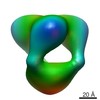 EMDB-5936: 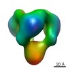 EMDB-5937:  EMDB-5938:  EMDB-5939: |
| Source |
|
 Moloney murine leukemia virus
