 Movie
Movie Controller
 Structure viewers
 About Yorodumi Papers


+Search query
-Structure paper
| Title | Domain organization of membrane-bound factor VIII. |
|---|---|
| Journal, issue, pages | Biopolymers, Vol. 99, Issue 7, Page 448-459, Year 2013 |
| Publish date | Apr 22, 2016 |
 Authors Authors | Svetla Stoilova-McPhie / Gillian C Lynch / Steven Ludtke / B Montgomery Pettitt /  |
| PubMed Abstract | Factor VIII (FVIII) is the blood coagulation protein which when defective or deficient causes for hemophilia A, a severe hereditary bleeding disorder. Activated FVIII (FVIIIa) is the cofactor to the ...Factor VIII (FVIII) is the blood coagulation protein which when defective or deficient causes for hemophilia A, a severe hereditary bleeding disorder. Activated FVIII (FVIIIa) is the cofactor to the serine protease factor IXa (FIXa) within the membrane-bound Tenase complex, responsible for amplifying its proteolytic activity more than 100,000 times, necessary for normal clot formation. FVIII is composed of two noncovalently linked peptide chains: a light chain (LC) holding the membrane interaction sites and a heavy chain (HC) holding the main FIXa interaction sites. The interplay between the light and heavy chains (HCs) in the membrane-bound state is critical for the biological efficiency of FVIII. Here, we present our cryo-electron microscopy (EM) and structure analysis studies of human FVIII-LC, when helically assembled onto negatively charged single lipid bilayer nanotubes. The resolved FVIII-LC membrane-bound structure supports aspects of our previously proposed FVIII structure from membrane-bound two-dimensional (2D) crystals, such as only the C2 domain interacts directly with the membrane. The LC is oriented differently in the FVIII membrane-bound helical and 2D crystal structures based on EM data, and the existing X-ray structures. This flexibility of the FVIII-LC domain organization in different states is discussed in the light of the FVIIIa-FIXa complex assembly and function. |
 External links External links | Biopolymers / PubMed:23616213 / PubMed Central |
| Methods | EM (helical sym.) / EM (electron crystallography) |
| Resolution | 15.0 Å |
| Structure data | 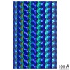 EMDB-5540: EMDB-5559: Cryo-em map of one molecule of factor VIII light chain from helically organized factor VIII light chain molecules bound to lipid nanotubes  PDB-3j2q: |
| Chemicals |  ChemComp-CU:  ChemComp-CA:  ChemComp-NAG: |
| Source |
|
 Keywords Keywords |  BLOOD CLOTTING / blood coagulation / cofactor / factor VIII / hemophilia / membrane binding BLOOD CLOTTING / blood coagulation / cofactor / factor VIII / hemophilia / membrane binding |


