 Movie
Movie Controller
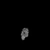
 Structure viewers
 About Yorodumi Papers


+Search query
-Structure paper
| Title | Molecular basis for the regulation of human phosphorylase kinase by phosphorylation and Ca. |
|---|---|
| Journal, issue, pages | Nat Commun, Vol. 16, Issue 1, Page 3020, Year 2025 |
| Publish date | Mar 28, 2025 |
 Authors Authors | Ruifang Ma / Bowen Du / Chen Shi / Lei Wang / Fuxing Zeng / Jie Han / Huiyi Guan / Yong Wang / Kaige Yan /  |
| PubMed Abstract | Phosphorylase kinase (PhK) regulates the degradation of glycogen by integrating diverse signals, providing energy to the organism. Dysfunctional mutations may directly lead to Glycogen Storage ...Phosphorylase kinase (PhK) regulates the degradation of glycogen by integrating diverse signals, providing energy to the organism. Dysfunctional mutations may directly lead to Glycogen Storage Disease type IX (GSD IX), whereas the abnormal expression of PhK is also associated with tumors. Here, we use cryo-electron microscopy (cryo-EM) to resolve its near-atomic structures in the inactive and active states. These structures reveal the interactions and relative locations of the four subunits (αβγδ) within the PhK complex. Phosphorylated α and β subunits induce PhK to present a more compact state, while Ca causes sliding of the δ subunit along the helix of the γ subunit. Both actions synergistically activate PhK by enabling the de-inhibition of the γ subunit. We also identified different binding modes between PhK and its substrate, glycogen phosphorylase (GP), in two distinct states, using cross-linking mass spectrometry (XL-MS). This study provides valuable insights into the regulatory mechanisms of PhK, thereby enhancing our understanding of GSD IX and its implications in tumorigenesis. |
 External links External links | Nat Commun / PubMed:40148320 / PubMed Central |
| Methods | EM (single particle) |
| Resolution | 3.23 - 4.24 Å |
| Structure data | EMDB-39775, PDB-8z5m: EMDB-39777, PDB-8z5p: EMDB-39778, PDB-8z5q: EMDB-39779, PDB-8z5r: EMDB-39781, PDB-8z5t:  EMDB-39784: human phosphorylase kinase alpha/beta/gamma/delta subcomplex - inactive state  EMDB-39785: human phosphorylase kinase alpha/beta/gamma/delta subcomplex - phosphorylation and Ca2+ bound state |
| Chemicals |  ChemComp-ATP: |
| Source |
|
 Keywords Keywords | CYTOSOLIC PROTEIN / Kinase / glycogenolysis / SIGNALING PROTEIN |


 homo sapiens (human)