Movie
Movie Controller
 Structure viewers
 About Yorodumi Papers


+Search query
-Structure paper
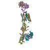
| Title | Structure and assembly of the dystrophin glycoprotein complex. |
|---|---|
| Journal, issue, pages | Nature, Vol. 637, Issue 8048, Page 1252-1260, Year 2025 |
| Publish date | Dec 11, 2024 |
 Authors Authors | Li Wan / Xiaofei Ge / Qikui Xu / Gaoxingyu Huang / Tiandi Yang / Kevin P Campbell / Zhen Yan / Jianping Wu /   |
| PubMed Abstract | The dystrophin glycoprotein complex (DGC) has a crucial role in maintaining cell membrane stability and integrity by connecting the intracellular cytoskeleton with the surrounding extracellular ...The dystrophin glycoprotein complex (DGC) has a crucial role in maintaining cell membrane stability and integrity by connecting the intracellular cytoskeleton with the surrounding extracellular matrix. Dysfunction of dystrophin and its associated proteins results in muscular dystrophy, a disorder characterized by progressive muscle weakness and degeneration. Despite the important roles of the DGC in physiology and pathology, its structural details remain largely unknown, hindering a comprehensive understanding of its assembly and function. Here we isolated the native DGC from mouse skeletal muscle and obtained its high-resolution structure. Our findings unveil a markedly divergent structure from the previous model of DGC assembly. Specifically, on the extracellular side, β-, γ- and δ-sarcoglycans co-fold to form a specialized, extracellular tower-like structure, which has a central role in complex assembly by providing binding sites for α-sarcoglycan and dystroglycan. In the transmembrane region, sarcoglycans and sarcospan flank and stabilize the single transmembrane helix of dystroglycan, rather than forming a subcomplex as previously proposed. On the intracellular side, sarcoglycans and dystroglycan engage in assembly with the dystrophin-dystrobrevin subcomplex through extensive interaction with the ZZ domain of dystrophin. Collectively, these findings enhance our understanding of the structural linkage across the cell membrane and provide a foundation for the molecular interpretation of many muscular dystrophy-related mutations. |
 External links External links | Nature / PubMed:39663450 / PubMed Central |
| Methods | EM (single particle) |
| Resolution | 3.2 - 3.5 Å |
| Structure data | EMDB-39568, PDB-8yt8:  EMDB-39569: Cryo-EM structure of the dystrophin glycoprotein complex |
| Chemicals |  ChemComp-NAG:  ChemComp-CLR:  ChemComp-ZN:  ChemComp-P5S:  ChemComp-CA: |
| Source |
|
 Keywords Keywords | STRUCTURAL PROTEIN / complex membrane stability signaling |