 Movie
Movie Controller
 Structure viewers
 About Yorodumi Papers


+Search query
-Structure paper
| Title | Single-particle cryoelectron microscopy analysis reveals the HIV-1 spike as a tripod structure. |
|---|---|
| Journal, issue, pages | Proc Natl Acad Sci U S A, Vol. 107, Issue 44, Page 18844-18849, Year 2010 |
| Publish date | Nov 2, 2010 |
 Authors Authors | Shang-Rung Wu / Robin Löving / Birgitta Lindqvist / Hans Hebert / Philip J B Koeck / Mathilda Sjöberg / Henrik Garoff /  |
| PubMed Abstract | The HIV-1 spike is a trimer of the transmembrane gp41 and the peripheral gp120 subunit pair. It is activated for virus-cell membrane fusion by binding sequentially to CD4 and to a chemokine receptor. ...The HIV-1 spike is a trimer of the transmembrane gp41 and the peripheral gp120 subunit pair. It is activated for virus-cell membrane fusion by binding sequentially to CD4 and to a chemokine receptor. Here we have studied the structural transition of the trimeric spike during the activation process. We solubilized and isolated unliganded and CD4-bound spikes from virus-like particles and used cryoelectron microscopy to reconstruct their 3D structures. In order to increase the yield and stability of the spike, we used an endodomain deleted and gp120-gp41 disulfide-linked variant. The unliganded spike displayed a hollow cage-like structure where the gp120-gp41 protomeric units formed a roof and bottom, and separated lobes and legs on the sides. The tripod structure was verified by fitting the recent atomic core structure of gp120 with intact N- and C-terminal ends into the spike density map. This defined the lobe as gp120 core, showed that the legs contained the polypeptide termini, and suggested the deleted variable loops V1/V2 and V3 to occupy the roof and gp41 the bottom. CD4 binding shifted the roof density peripherally and condensed the bottom density centrally. Fitting with a V3 containing gp120 core suggested that the V1/V2 loops in the roof were displaced laterally and the V3 lifted up, while the core and leg were kept in place. The loop displacements probably prepared the spike for coreceptor interaction and roof opening so that a new fusion-active gp41 structure, assembled at the center of the cage bottom, could reach the target membrane. |
 External links External links | Proc Natl Acad Sci U S A / PubMed:20956336 / PubMed Central |
| Methods | EM (single particle) |
| Resolution | 18.0 - 21.0 Å |
| Structure data | 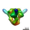 EMDB-1800:  EMDB-1801: |
| Source |
|

 Human immunodeficiency virus 1
 Homo sapiens (human)