 Movie
Movie Controller
 Structure viewers
 About Yorodumi Papers


+Search query
-Structure paper
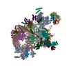
| Title | Structural basis of catalytic activation in human splicing. |
|---|---|
| Journal, issue, pages | Nature, Vol. 617, Issue 7962, Page 842-850, Year 2023 |
| Publish date | May 10, 2023 |
 Authors Authors | Jana Schmitzová / Constantin Cretu / Christian Dienemann / Henning Urlaub / Vladimir Pena /   |
| PubMed Abstract | Pre-mRNA splicing follows a pathway driven by ATP-dependent RNA helicases. A crucial event of the splicing pathway is the catalytic activation, which takes place at the transition between the ...Pre-mRNA splicing follows a pathway driven by ATP-dependent RNA helicases. A crucial event of the splicing pathway is the catalytic activation, which takes place at the transition between the activated B and the branching-competent B spliceosomes. Catalytic activation occurs through an ATP-dependent remodelling mediated by the helicase PRP2 (also known as DHX16). However, because PRP2 is observed only at the periphery of spliceosomes, its function has remained elusive. Here we show that catalytic activation occurs in two ATP-dependent stages driven by two helicases: PRP2 and Aquarius. The role of Aquarius in splicing has been enigmatic. Here the inactivation of Aquarius leads to the stalling of a spliceosome intermediate-the B complex-found halfway through the catalytic activation process. The cryogenic electron microscopy structure of B reveals how PRP2 and Aquarius remodel B and B, respectively. Notably, PRP2 translocates along the intron while it strips away the RES complex, opens the SF3B1 clamp and unfastens the branch helix. Translocation terminates six nucleotides downstream of the branch site through an assembly of PPIL4, SKIP and the amino-terminal domain of PRP2. Finally, Aquarius enables the dissociation of PRP2, plus the SF3A and SF3B complexes, which promotes the relocation of the branch duplex for catalysis. This work elucidates catalytic activation in human splicing, reveals how a DEAH helicase operates and provides a paradigm for how helicases can coordinate their activities. |
 External links External links | Nature / PubMed:37165190 / PubMed Central |
| Methods | EM (single particle) |
| Resolution | 3.1 - 5.9 Å |
| Structure data | EMDB-14146, PDB-7qtt: EMDB-16658, PDB-8ch6: |
| Chemicals |  ChemComp-ZN:  ChemComp-IHP:  ChemComp-GTP:  ChemComp-MG: |
| Source |
|
 Keywords Keywords | SPLICING / Spliceosome / helicase / Prp2 / Aquarius / catalytic activation / activated spliceosome |


 homo sapiens (human)
 unidentified adenovirus