[English] 日本語
Yorodumi Papers
- Database of articles cited by EMDB/PDB/SASBDB data -

+
Search query

Keywords
Structure methods
Author
Journal
IF

-
Structure paper

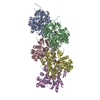
TitleStructural basis of actin filament assembly and aging.
Journal, issue, pagesNature, Vol. 611, Issue 7935, Page 374-379, Year 2022
Publish dateOct 26, 2022
AuthorsWout Oosterheert / Björn U Klink / Alexander Belyy / Sabrina Pospich / Stefan Raunser /
PubMed AbstractThe dynamic turnover of actin filaments (F-actin) controls cellular motility in eukaryotes and is coupled to changes in the F-actin nucleotide state. It remains unclear how F-actin hydrolyses ATP and ...The dynamic turnover of actin filaments (F-actin) controls cellular motility in eukaryotes and is coupled to changes in the F-actin nucleotide state. It remains unclear how F-actin hydrolyses ATP and subsequently undergoes subtle conformational rearrangements that ultimately lead to filament depolymerization by actin-binding proteins. Here we present cryo-electron microscopy structures of F-actin in all nucleotide states, polymerized in the presence of Mg or Ca at approximately 2.2 Å resolution. The structures show that actin polymerization induces the relocation of water molecules in the nucleotide-binding pocket, activating one of them for the nucleophilic attack of ATP. Unexpectedly, the back door for the subsequent release of inorganic phosphate (P) is closed in all structures, indicating that P release occurs transiently. The small changes in the nucleotide-binding pocket after ATP hydrolysis and P release are sensed by a key amino acid, amplified and transmitted to the filament periphery. Furthermore, differences in the positions of water molecules in the nucleotide-binding pocket explain why Ca-actin shows slower polymerization rates than Mg-actin. Our work elucidates the solvent-driven rearrangements that govern actin filament assembly and aging and lays the foundation for the rational design of drugs and small molecules for imaging and therapeutic applications.
External linksNature / PubMed:36289337 / PubMed Central
MethodsEM (single particle)
Resolution2.15 - 2.24 Å
Structure data

EMDB-15104, PDB-8a2r:
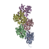
Cryo-EM structure of F-actin in the Mg2+-ADP-BeF3- nucleotide state.
Method: EM (single particle) / Resolution: 2.17 Å

EMDB-15105, PDB-8a2s:
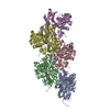
Cryo-EM structure of F-actin in the Mg2+-ADP-Pi nucleotide state.
Method: EM (single particle) / Resolution: 2.22 Å

EMDB-15106, PDB-8a2t:
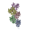
Cryo-EM structure of F-actin in the Mg2+-ADP nucleotide state.
Method: EM (single particle) / Resolution: 2.24 Å

EMDB-15107, PDB-8a2u:
Cryo-EM structure of F-actin in the Ca2+-ADP-BeF3- nucleotide state.
Method: EM (single particle) / Resolution: 2.21 Å

EMDB-15108, PDB-8a2y:
Cryo-EM structure of F-actin in the Ca2+-ADP-Pi nucleotide state.
Method: EM (single particle) / Resolution: 2.15 Å

EMDB-15109, PDB-8a2z:
Cryo-EM structure of F-actin in the Ca2+-ADP nucleotide state.
Method: EM (single particle) / Resolution: 2.15 Å

Chemicals

ChemComp-ADP:
ADENOSINE-5'-DIPHOSPHATE / ADP, energy-carrying molecule*YM

ChemComp-MG:
Unknown entry

ChemComp-BEF:
BERYLLIUM TRIFLUORIDE ION

ChemComp-HOH:
WATER

ChemComp-PO4:
PHOSPHATE ION

ChemComp-CA:
Unknown entry

Source
  • oryctolagus cuniculus (rabbit)
  • rabbit (rabbit)
KeywordsSTRUCTURAL PROTEIN / actin / cytoskeleton / filament / nucleotide state

+
About Yorodumi Papers

-
News

-
Feb 9, 2022. New format data for meta-information of EMDB entries

New format data for meta-information of EMDB entries

  • Version 3 of the EMDB header file is now the official format.
  • The previous official version 1.9 will be removed from the archive.

Related info.:EMDB header

External links:wwPDB to switch to version 3 of the EMDB data model

-
Aug 12, 2020. Covid-19 info

Covid-19 info

URL: https://pdbj.org/emnavi/covid19.php

New page: Covid-19 featured information page in EM Navigator.

Related info.:Covid-19 info / Mar 5, 2020. Novel coronavirus structure data

+
Mar 5, 2020. Novel coronavirus structure data

Novel coronavirus structure data

Related info.:Yorodumi Speices / Aug 12, 2020. Covid-19 info

External links:COVID-19 featured content - PDBj / Molecule of the Month (242):Coronavirus Proteases

+
Jan 31, 2019. EMDB accession codes are about to change! (news from PDBe EMDB page)

EMDB accession codes are about to change! (news from PDBe EMDB page)

  • The allocation of 4 digits for EMDB accession codes will soon come to an end. Whilst these codes will remain in use, new EMDB accession codes will include an additional digit and will expand incrementally as the available range of codes is exhausted. The current 4-digit format prefixed with “EMD-” (i.e. EMD-XXXX) will advance to a 5-digit format (i.e. EMD-XXXXX), and so on. It is currently estimated that the 4-digit codes will be depleted around Spring 2019, at which point the 5-digit format will come into force.
  • The EM Navigator/Yorodumi systems omit the EMD- prefix.

Related info.:Q: What is EMD? / ID/Accession-code notation in Yorodumi/EM Navigator

External links:EMDB Accession Codes are Changing Soon! / Contact to PDBj

+
Jul 12, 2017. Major update of PDB

Major update of PDB

  • wwPDB released updated PDB data conforming to the new PDBx/mmCIF dictionary.
  • This is a major update changing the version number from 4 to 5, and with Remediation, in which all the entries are updated.
  • In this update, many items about electron microscopy experimental information are reorganized (e.g. em_software).
  • Now, EM Navigator and Yorodumi are based on the updated data.

External links:wwPDB Remediation / Enriched Model Files Conforming to OneDep Data Standards Now Available in the PDB FTP Archive

-
Yorodumi Papers

Database of articles cited by EMDB/PDB/SASBDB data

  • Database of articles cited by EMDB, PDB, and SASBDB entries
  • Using PubMed data

Related info.:EMDB / PDB / SASBDB / Yorodumi / EMN Papers / Changes in new EM Navigator and Yorodumi

Read more