Jun Liu / Dianne W Taylor / Elena B Krementsova / Kathleen M Trybus / Kenneth A Taylor /
PubMed Abstract
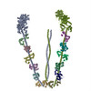
Unconventional myosin V (myoV) is an actin-based molecular motor that has a key function in organelle and mRNA transport, as well as in membrane trafficking. MyoV was the first member of the myosin ...Unconventional myosin V (myoV) is an actin-based molecular motor that has a key function in organelle and mRNA transport, as well as in membrane trafficking. MyoV was the first member of the myosin superfamily shown to be processive, meaning that a single motor protein can 'walk' hand-over-hand along an actin filament for many steps before detaching. Full-length myoV has a low actin-activated MgATPase activity at low [Ca2+], whereas expressed constructs lacking the cargo-binding domain have a high activity regardless of [Ca2+] (refs 5-7). Hydrodynamic data and electron micrographs indicate that the active state is extended, whereas the inactive state is compact. Here we show the first three-dimensional structure of the myoV inactive state. Each myoV molecule consists of two heads that contain an amino-terminal motor domain followed by a lever arm that binds six calmodulins. The heads are followed by a coiled-coil dimerization domain (S2) and a carboxy-terminal globular cargo-binding domain. In the inactive structure, bending of myoV at the head-S2 junction places the cargo-binding domain near the motor domain's ATP-binding pocket, indicating that ATPase inhibition might occur through decreased rates of nucleotide exchange. The actin-binding interfaces are unobstructed, and the lever arm is oriented in a position typical of strong actin-binding states. This structure indicates that motor recycling after cargo delivery might occur through transport on actively treadmilling actin filaments rather than by diffusion.
EMDB-1201: Three-dimensional structure of the myosin V inhibited state by cryoelectron tomography. PDB-2dfs: 3-D structure of Myosin-V inhibited state Method: EM (subtomogram averaging) / Resolution: 24.0 Å
Source
mus musculus (house mouse)
gallus gallus (chicken)
Keywords
CONTRACTILE PROTEIN/TRANSPORT PROTEIN / myosin-V / inhibited state / calmodulin / cryoelectron tomography / CONTRACTILE PROTEIN-TRANSPORT PROTEIN COMPLEX
+
About Yorodumi Papers
-
News
-
Feb 9, 2022. New format data for meta-information of EMDB entries
New format data for meta-information of EMDB entries
Version 3 of the EMDB header file is now the official format.
The previous official version 1.9 will be removed from the archive.
In the structure databanks used in Yorodumi, some data are registered as the other names, "COVID-19 virus" and "2019-nCoV". Here are the details of the virus and the list of structure data.
Jan 31, 2019. EMDB accession codes are about to change! (news from PDBe EMDB page)
EMDB accession codes are about to change! (news from PDBe EMDB page)
The allocation of 4 digits for EMDB accession codes will soon come to an end. Whilst these codes will remain in use, new EMDB accession codes will include an additional digit and will expand incrementally as the available range of codes is exhausted. The current 4-digit format prefixed with “EMD-” (i.e. EMD-XXXX) will advance to a 5-digit format (i.e. EMD-XXXXX), and so on. It is currently estimated that the 4-digit codes will be depleted around Spring 2019, at which point the 5-digit format will come into force.
The EM Navigator/Yorodumi systems omit the EMD- prefix.
Related info.:Q: What is EMD? / ID/Accession-code notation in Yorodumi/EM Navigator
 Movie
Movie Controller
 Structure viewers
 About Yorodumi Papers


 Authors
 External links
 Keywords