 Movie
Movie Controller
 Structure viewers
 About Yorodumi Papers


+Search query
-Structure paper
| Title | Structural analysis of pathogenic mutations targeting Glu427 of ALDH7A1, the hot spot residue of pyridoxine-dependent epilepsy. |
|---|---|
| Journal, issue, pages | J Inherit Metab Dis, Vol. 43, Issue 3, Page 635-644, Year 2020 |
| Publish date | Dec 1, 2019 |
 Authors Authors | Adrian R Laciak / David A Korasick / Kent S Gates / John J Tanner /  |
| PubMed Abstract | Certain loss-of-function mutations in the gene encoding the lysine catabolic enzyme aldehyde dehydrogenase 7A1 (ALDH7A1) cause pyridoxine-dependent epilepsy (PDE). Missense mutations of Glu427, ...Certain loss-of-function mutations in the gene encoding the lysine catabolic enzyme aldehyde dehydrogenase 7A1 (ALDH7A1) cause pyridoxine-dependent epilepsy (PDE). Missense mutations of Glu427, especially Glu427Gln, account for ~30% of the mutated alleles in PDE patients, and thus Glu427 has been referred to as a mutation hot spot of PDE. Glu427 is invariant in the ALDH superfamily and forms ionic hydrogen bonds with the nicotinamide ribose of the NAD cofactor. Here we report the first crystal structures of ALDH7A1 containing pathogenic mutations targeting Glu427. The mutant enzymes E427Q, Glu427Asp, and Glu427Gly were expressed in Escherichia coli and purified. The recombinant enzymes displayed negligible catalytic activity compared to the wild-type enzyme. The crystal structures of the mutant enzymes complexed with NAD were determined to understand how the mutations impact NAD binding. In the E427Q and E427G structures, the nicotinamide mononucleotide is highly flexible and lacks a defined binding pose. In E427D, the bound NAD adopts a "retracted" conformation in which the nicotinamide ring is too far from the catalytic Cys residue for hydride transfer. Thus, the structures revealed a shared mechanism for loss of function: none of the variants are able to stabilise the nicotinamide of NAD in the pose required for catalysis. We also show that these mutations reduce the amount of active tetrameric ALDH7A1 at the concentration of NAD tested. Altogether, our results provide the three-dimensional molecular structural basis of the most common pathogenic variants of PDE and implicate strong (ionic) hydrogen bonds in the aetiology of a human disease. |
 External links External links | J Inherit Metab Dis / PubMed:31652343 / PubMed Central |
| Methods | SAS (X-ray synchrotron) / X-ray diffraction |
| Resolution | 1.75 - 2.15 Å |
| Structure data | 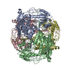 SASDGH4:  SASDGJ4:  SASDGK4: 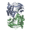 SASDGL4:  SASDGM4:  SASDGN4:  SASDGP4:  SASDGQ4:  SASDGR4:  SASDGS4:  SASDGT4:  SASDGU4:  PDB-6o4i: 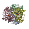 PDB-6o4k: 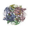 PDB-6o4l: 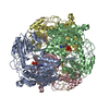 PDB-6u2x: |
| Chemicals | 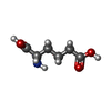 ChemComp-UN1:  ChemComp-EDO:  ChemComp-HOH:  ChemComp-NAD:  ChemComp-CL: |
| Source |
|
 Keywords Keywords | OXIDOREDUCTASE / ALDEHYDE DEHYDROGENASE / NAD / LYSINE CATABOLISM |
 homo sapiens (human)