ムービー
ムービー コントローラー
 構造ビューア
 万見文献について


+検索条件
-Structure paper
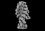
| タイトル | Double-headed binding of myosin II to F-actin shows the effect of strain on head structure. |
|---|---|
| ジャーナル・号・ページ | J Struct Biol, Vol. 215, Issue 3, Page 107995, Year 2023 |
| 掲載日 | 2023年7月4日 |
 著者 著者 | Alimohammad Hojjatian / Dianne W Taylor / Nadia Daneshparvar / Patricia M Fagnant / Kathleen M Trybus / Kenneth A Taylor /  |
| PubMed 要旨 | Force production in muscle is achieved through the interaction of myosin and actin. Strong binding states in active muscle are associated with Mg·ADP bound to the active site; release of Mg·ADP ...Force production in muscle is achieved through the interaction of myosin and actin. Strong binding states in active muscle are associated with Mg·ADP bound to the active site; release of Mg·ADP allows rebinding of ATP and dissociation from actin. Thus, Mg·ADP binding is positioned for adaptation as a force sensor. Mechanical loads on the lever arm can affect the ability of myosin to release Mg·ADP but exactly how this is done is poorly defined. Here we use F-actin decorated with double-headed smooth muscle myosin fragments in the presence of Mg·ADP to visualize the effect of internally supplied tension on the paired lever arms using cryoEM. The interaction of the paired heads with two adjacent actin subunits is predicted to place one lever arm under positive and the other under negative strain. The converter domain is believed to be the most flexible domain within myosin head. Our results, instead, point to the segment of heavy chain between the essential and regulatory light chains as the location of the largest structural change. Moreover, our results suggest no large changes in the myosin coiled coil tail as the locus of strain relief when both heads bind F-actin. The method would be adaptable to double-headed members of the myosin family. We anticipate that the study of actin-myosin interaction using double-headed fragments enables visualization of domains that are typically noisy in decoration with single-headed fragments. |
 リンク リンク | J Struct Biol / PubMed:37414375 / PubMed Central |
| 手法 | EM (単粒子) |
| 解像度 | 19.0 Å |
| 構造データ | EMDB-29646: Acto-HMM complex in ADP-state. Chicken smooth muscle HMM and chicken pectoralis actin |
| 由来 |
|
 キーワード キーワード | MOTOR PROTEIN / Actin / Myosin / Smooth muscle / Cryo-EM / Cryo-ET / Heavy meromyosin |