 Movie
Movie Controller

[English] 日本語
 Yorodumi
- PDB-8syf: Homology model of Acto-HMM complex in ADP-state. Chicken smooth m... -
+ Open data

- Basic information
| Entry | Database: PDB / ID: 8syf | ||||||||||||||||||||||||||||||
|---|---|---|---|---|---|---|---|---|---|---|---|---|---|---|---|---|---|---|---|---|---|---|---|---|---|---|---|---|---|---|---|
| Title | Homology model of Acto-HMM complex in ADP-state. Chicken smooth muscle HMM and chicken pectoralis actin | ||||||||||||||||||||||||||||||
 Components Components |
| ||||||||||||||||||||||||||||||
 Keywords Keywords | MOTOR PROTEIN / Actin / Myosin / Smooth muscle / Cryo-EM / Cryo-ET / Heavy meromyosin | ||||||||||||||||||||||||||||||
| Function / homology |  Function and homology information Function and homology informationRHO GTPases activate PAKs / myosin II filament / Smooth Muscle Contraction / myofibril assembly / myosin II binding / muscle myosin complex / actomyosin / actomyosin structure organization / myosin complex / myosin II complex ...RHO GTPases activate PAKs / myosin II filament / Smooth Muscle Contraction / myofibril assembly / myosin II binding / muscle myosin complex / actomyosin / actomyosin structure organization / myosin complex / myosin II complex / structural constituent of muscle / microfilament motor activity / myosin heavy chain binding / myofibril / cytoskeletal motor activity / striated muscle thin filament / skeletal muscle thin filament assembly / skeletal muscle fiber development / stress fiber / actin filament / ADP binding / Hydrolases; Acting on acid anhydrides; Acting on acid anhydrides to facilitate cellular and subcellular movement / structural constituent of cytoskeleton / actin filament binding / actin cytoskeleton / hydrolase activity / calcium ion binding / magnesium ion binding / ATP binding / cytosol / cytoplasm Similarity search - Function | ||||||||||||||||||||||||||||||
| Biological species |  | ||||||||||||||||||||||||||||||
| Method | ELECTRON MICROSCOPY / single particle reconstruction / cryo EM / Resolution: 19 Å | ||||||||||||||||||||||||||||||
 Authors Authors | Hojjatian, A. / Taylor, D.W. / Daneshparvar, N. / Trybus, K.M. / Taylor, K.A. | ||||||||||||||||||||||||||||||
| Funding support |  United States, 9items United States, 9items
| ||||||||||||||||||||||||||||||
 Citation Citation | Journal: J Struct Biol / Year: 2023 Title: Double-headed binding of myosin II to F-actin shows the effect of strain on head structure. Authors: Alimohammad Hojjatian / Dianne W Taylor / Nadia Daneshparvar / Patricia M Fagnant / Kathleen M Trybus / Kenneth A Taylor / Abstract: Force production in muscle is achieved through the interaction of myosin and actin. Strong binding states in active muscle are associated with Mg·ADP bound to the active site; release of Mg·ADP ...Force production in muscle is achieved through the interaction of myosin and actin. Strong binding states in active muscle are associated with Mg·ADP bound to the active site; release of Mg·ADP allows rebinding of ATP and dissociation from actin. Thus, Mg·ADP binding is positioned for adaptation as a force sensor. Mechanical loads on the lever arm can affect the ability of myosin to release Mg·ADP but exactly how this is done is poorly defined. Here we use F-actin decorated with double-headed smooth muscle myosin fragments in the presence of Mg·ADP to visualize the effect of internally supplied tension on the paired lever arms using cryoEM. The interaction of the paired heads with two adjacent actin subunits is predicted to place one lever arm under positive and the other under negative strain. The converter domain is believed to be the most flexible domain within myosin head. Our results, instead, point to the segment of heavy chain between the essential and regulatory light chains as the location of the largest structural change. Moreover, our results suggest no large changes in the myosin coiled coil tail as the locus of strain relief when both heads bind F-actin. The method would be adaptable to double-headed members of the myosin family. We anticipate that the study of actin-myosin interaction using double-headed fragments enables visualization of domains that are typically noisy in decoration with single-headed fragments. | ||||||||||||||||||||||||||||||
| History |
|
- Structure visualization
| Structure viewer | Molecule: MolmilJmol/JSmol |
|---|
- Downloads & links
-Download
| PDBx/mmCIF format | 8syf.cif.gz | 515.6 KB | Display | PDBx/mmCIF format |
|---|---|---|---|---|
| PDB format | pdb8syf.ent.gz | 415.7 KB | Display | PDB format |
| PDBx/mmJSON format | 8syf.json.gz | Tree view | PDBx/mmJSON format | |
| Others |  Other downloads Other downloads |
-Validation report
| Arichive directory | https://data.pdbj.org/pub/pdb/validation_reports/sy/8syfftp://data.pdbj.org/pub/pdb/validation_reports/sy/8syf | HTTPS FTP |
|---|
-Related structure data
| Related structure data | 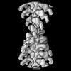 29646MC M: map data used to model this data C: citing same article ( |
|---|---|
| Similar structure data | |
| Experimental dataset #1 | Data reference: 10.6019/EMPIAR-11555 / Data set type: EMPIAR |
-Links
 PDBj
PDBj


- Assembly
| Deposited unit | 
|
|---|---|
| 1 |
|
-Components
| #1: Protein | Mass: 96768.078 Da / Num. of mol.: 2 Source method: isolated from a genetically manipulated source Source: (gene. exp.)  unidentified baculovirus / References: UniProt: A0A8V0ZE13 unidentified baculovirus / References: UniProt: A0A8V0ZE13#2: Protein | Mass: 41862.613 Da / Num. of mol.: 2 / Source method: isolated from a natural source / Source: (natural) References: UniProt: P68139, Hydrolases; Acting on acid anhydrides; Acting on acid anhydrides to facilitate cellular and subcellular movement #3: Protein | Mass: 16639.715 Da / Num. of mol.: 2 Source method: isolated from a genetically manipulated source Source: (gene. exp.) unidentified baculovirus / References: UniProt: P02607#4: Protein | Mass: 16583.412 Da / Num. of mol.: 2 Source method: isolated from a genetically manipulated source Source: (gene. exp.) unidentified baculovirus / References: UniProt: P02612Has protein modification | N | |
|---|
-Experimental details
-Experiment
| Experiment | Method: ELECTRON MICROSCOPY |
|---|---|
| EM experiment | Aggregation state: FILAMENT / 3D reconstruction method: single particle reconstruction |
- Sample preparation
| Component | Name: Filamentous actin decorated with chicken smooth muscle heavy meromyosin Type: COMPLEX / Entity ID: all / Source: RECOMBINANT |
|---|---|
| Molecular weight | Experimental value: NO |
| Source (natural) | Organism: |
| Source (recombinant) | Organism: unidentified baculovirus |
| Buffer solution | pH: 7.6 |
| Specimen | Embedding applied: NO / Shadowing applied: NO / Staining applied: NO / Vitrification applied: YES |
| Vitrification | Cryogen name: ETHANE-PROPANE |
- Electron microscopy imaging
| Experimental equipment |  Model: Titan Krios / Image courtesy: FEI Company |
|---|---|
| Microscopy | Model: FEI TITAN KRIOS |
| Electron gun | Electron source:  FIELD EMISSION GUN / Accelerating voltage: 300 kV / Illumination mode: OTHER FIELD EMISSION GUN / Accelerating voltage: 300 kV / Illumination mode: OTHER |
| Electron lens | Mode: BRIGHT FIELD / Nominal defocus max: 2000 nm / Nominal defocus min: 200 nm |
| Image recording | Electron dose: 29.6 e/Å2 / Detector mode: INTEGRATING / Film or detector model: DIRECT ELECTRON DE-64 (8k x 8k) |
- Processing
| EM software | Name: PHENIX / Category: model refinement | ||||||||||||||||||||||||
|---|---|---|---|---|---|---|---|---|---|---|---|---|---|---|---|---|---|---|---|---|---|---|---|---|---|
| CTF correction | Type: NONE | ||||||||||||||||||||||||
| 3D reconstruction | Resolution: 19 Å / Resolution method: FSC 0.143 CUT-OFF / Num. of particles: 17394 / Symmetry type: POINT | ||||||||||||||||||||||||
| Refine LS restraints |
|
