 ムービー
ムービー コントローラー
 構造ビューア
 万見文献について


+検索条件
-Structure paper
| タイトル | BAK core dimers bind lipids and can be bridged by them. |
|---|---|
| ジャーナル・号・ページ | Nat. Struct. Mol. Biol., Vol. 27, Page 1024-1031, Year 2020 |
| 掲載日 | 2019年11月7日 (構造データの登録日) |
 著者 著者 | Cowan, A.D. / Smith, N.A. / Sandow, J.J. / Kapp, E.A. / Rustam, Y.H. / Murphy, J.M. / Brouwer, J.M. / Bernardini, J.P. / Roy, M.J. / Wardak, A.Z. ...Cowan, A.D. / Smith, N.A. / Sandow, J.J. / Kapp, E.A. / Rustam, Y.H. / Murphy, J.M. / Brouwer, J.M. / Bernardini, J.P. / Roy, M.J. / Wardak, A.Z. / Tan, I.K. / Webb, A.I. / Gulbis, J.M. / Smith, B.J. / Reid, G.E. / Dewson, G. / Colman, P.M. / Czabotar, P.E. |
 リンク リンク | Nat. Struct. Mol. Biol. / PubMed:32929280 |
| 手法 | X線回折 |
| 解像度 | 1.696 - 2.492 Å |
| 構造データ | 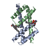 PDB-6uxm:  PDB-6uxn: 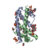 PDB-6uxo: 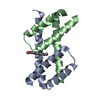 PDB-6uxp:  PDB-6uxq:  PDB-6uxr: |
| 化合物 |  ChemComp-PEE:  ChemComp-8SP:  ChemComp-SO4:  ChemComp-GOL:  ChemComp-HOH:  ChemComp-LMT:  ChemComp-ACT:  ChemComp-EDO:  ChemComp-PG8:  ChemComp-C8E:  ChemComp-LBN:  ChemComp-K6G:  ChemComp-PG4:  ChemComp-PGE: |
| 由来 |
|
 キーワード キーワード | APOPTOSIS / Pore-forming Protein |
 homo sapiens (ヒト)