ムービー
ムービー コントローラー
 構造ビューア
 万見文献について


+検索条件
-Structure paper
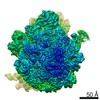
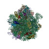
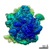
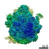
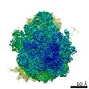
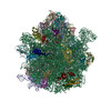
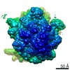
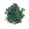
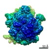
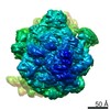
| タイトル | The structural basis for release-factor activation during translation termination revealed by time-resolved cryogenic electron microscopy. |
|---|---|
| ジャーナル・号・ページ | Nat Commun, Vol. 10, Issue 1, Page 2579, Year 2019 |
| 掲載日 | 2019年6月12日 |
 著者 著者 | Ziao Fu / Gabriele Indrisiunaite / Sandip Kaledhonkar / Binita Shah / Ming Sun / Bo Chen / Robert A Grassucci / Måns Ehrenberg / Joachim Frank /   |
| PubMed 要旨 | When the ribosome encounters a stop codon, it recruits a release factor (RF) to hydrolyze the ester bond between the peptide chain and tRNA. RFs have structural motifs that recognize stop codons in ...When the ribosome encounters a stop codon, it recruits a release factor (RF) to hydrolyze the ester bond between the peptide chain and tRNA. RFs have structural motifs that recognize stop codons in the decoding center and a GGQ motif for induction of hydrolysis in the peptidyl transfer center 70 Å away. Surprisingly, free RF2 is compact, with only 20 Å between its codon-reading and GGQ motifs. Cryo-EM showed that ribosome-bound RFs have extended structures, suggesting that RFs are compact when entering the ribosome and then extend their structures upon stop codon recognition. Here we use time-resolved cryo-EM to visualize transient compact forms of RF1 and RF2 at 3.5 and 4 Å resolution, respectively, in the codon-recognizing ribosome complex on the native pathway. About 25% of complexes have RFs in the compact state at 24 ms reaction time, and within 60 ms virtually all ribosome-bound RFs are transformed to their extended forms. |
 リンク リンク | Nat Commun / PubMed:31189921 / PubMed Central |
| 手法 | EM (単粒子) |
| 解像度 | 2.9 - 4.2 Å |
| 構造データ | EMDB-20173, PDB-6ore: EMDB-20174, PDB-6orl: EMDB-20184, PDB-6osk: EMDB-20187, PDB-6osq: EMDB-20188, PDB-6ost: EMDB-20193, PDB-6ot3: EMDB-20204, PDB-6ouo: |
| 化合物 |  ChemComp-MG:  ChemComp-ZN: |
| 由来 |
|
 キーワード キーワード | RIBOSOME / Time-resolved cryo-EM / Termination / short-lived / millisecond |