| タイトル | Structural determinants of protocadherin-15 mechanics and function in hearing and balance perception. |
|---|
| ジャーナル・号・ページ | Proc. Natl. Acad. Sci. USA, Year 2020 |
|---|
| 掲載日 | 2016年10月20日 (構造データの登録日) |
|---|
 著者 著者 | Choudhary, D. / Narui, Y. / Neel, B.L. / Wimalasena, L.N. / Klanseck, C.F. / De-la-Torre, P. / Chen, C. / Araya-Secchi, R. / Tamilselvan, E. / Sotomayor, M. |
|---|
 リンク リンク |  Proc. Natl. Acad. Sci. USA / Proc. Natl. Acad. Sci. USA /  PubMed:32963095 PubMed:32963095 |
|---|
| 手法 | X線回折 |
|---|
| 解像度 | 2 - 3.79 Å |
|---|
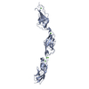
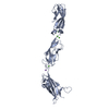
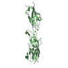
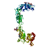
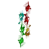
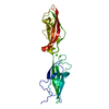
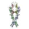
| 構造データ | PDB-5tpk:
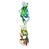
Crystal Structure of Mouse Protocadherin-15 EC7-8 V875A
手法: X-RAY DIFFRACTION / 解像度: 2 Å PDB-5uly:
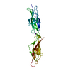
Crystal Structure of Human Protocadherin-15 EC2-3
手法: X-RAY DIFFRACTION / 解像度: 2.64 Å PDB-5w1d:
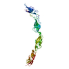
Crystal Structure of Mouse Protocadherin-15 EC4-7
手法: X-RAY DIFFRACTION / 解像度: 3.35 Å PDB-6bwn:
Crystal Structure of Mouse Protocadherin-15 EC6-7
手法: X-RAY DIFFRACTION / 解像度: 2.94 Å PDB-6bxu:
Crystal Structure of Mouse Protocadherin-15 EC5-7 I582T
手法: X-RAY DIFFRACTION / 解像度: 3.79 Å PDB-6e8f:
Crystal Structure of Human Protocadherin-15 EC3-5 CD2-1
手法: X-RAY DIFFRACTION / 解像度: 2.99 Å PDB-6eb5:
Crystal Structure of Human Protocadherin-15 EC2-3 V250N
手法: X-RAY DIFFRACTION / 解像度: 2.6 Å PDB-6eet:
Crystal structure of mouse Protocadherin-15 EC9-MAD12
手法: X-RAY DIFFRACTION / 解像度: 3.23 Å PDB-6mfo:
Crystal Structure of Human Protocadherin-15 EC1-3 G16D N369D Q370N
手法: X-RAY DIFFRACTION / 解像度: 3.15 Å PDB-6n22:
Crystal structure of mouse Protocadherin-15 EC1-2 BAP
手法: X-RAY DIFFRACTION / 解像度: 2.4 Å PDB-6n2e:
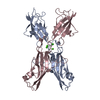
Crystal Structure of Human Protocadherin-15 EC1-3 G16D N369D Q370N and Mouse Cadherin-23 EC1-2 T15E
手法: X-RAY DIFFRACTION / 解像度: 2.9 Å |
|---|
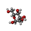
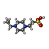
| 化合物 | ChemComp-EPE:
4-(2-HYDROXYETHYL)-1-PIPERAZINE ETHANESULFONIC ACID / HEPES / pH緩衝剤*YM
|
|---|
| 由来 |   mus musculus (ハツカネズミ) mus musculus (ハツカネズミ) homo sapiens (ヒト) homo sapiens (ヒト)
|
|---|
 キーワード キーワード | CELL ADHESION / hearing / mechanotransduction / adhesion / calcium-binding protein / stereocilia / hair cell / tip link / calcium binding protein |
|---|
 著者
著者 リンク
リンク Proc. Natl. Acad. Sci. USA /
  PubMed:32963095


 キーワード
キーワード ムービー
ムービー コントローラー
 構造ビューア
 万見文献について


 homo sapiens (ヒト)