 ムービー
ムービー コントローラー
 構造ビューア
 万見文献について


+検索条件
-Structure paper
| タイトル | The modular structure of the inner-membrane ring component PrgK facilitates assembly of the type III secretion system basal body. |
|---|---|
| ジャーナル・号・ページ | Structure, Vol. 23, Issue 1, Page 161-172, Year 2015 |
| 掲載日 | 2015年1月6日 |
 著者 著者 | Julien R C Bergeron / Liam J Worrall / Soumya De / Nikolaos G Sgourakis / Adrienne H Cheung / Emilie Lameignere / Mark Okon / Gregory A Wasney / David Baker / Lawrence P McIntosh / Natalie C J Strynadka /   |
| PubMed 要旨 | The type III secretion system (T3SS) is a large macromolecular assembly found at the surface of many pathogenic Gram-negative bacteria. Its role is to inject toxic "effector" proteins into the cells ...The type III secretion system (T3SS) is a large macromolecular assembly found at the surface of many pathogenic Gram-negative bacteria. Its role is to inject toxic "effector" proteins into the cells of infected organisms. The molecular details of the assembly of this large, multimembrane-spanning complex remain poorly understood. Here, we report structural, biochemical, and functional analyses of PrgK, an inner-membrane component of the prototypical Salmonella typhimurium T3SS. We have obtained the atomic structures of the two ring building globular domains and show that the C-terminal transmembrane helix is not essential for assembly and secretion. We also demonstrate that structural rearrangement of the two PrgK globular domains, driven by an interconnecting linker region, may promote oligomerization into ring structures. Finally, we used electron microscopy-guided symmetry modeling to propose a structural model for the intimately associated PrgH-PrgK ring interaction within the assembled basal body. |
 リンク リンク | Structure / PubMed:25533490 |
| 手法 | NMR (溶液) / EM (単粒子) / X線回折 |
| 解像度 | 2.6 - 11.7 Å |
| 構造データ |  PDB-2mky: 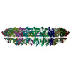 PDB-3j6d: 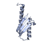 PDB-4oyc: 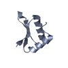 PDB-4w4m: |
| 化合物 |  ChemComp-HOH: |
| 由来 |
|
 キーワード キーワード | CELL INVASION / Secretion systems / macromolecular assemblies / STRUCTURAL PROTEIN / Bacterial secression macromolecular assemblies / PROTEIN TRANSPORT / T3SS / macromolecular assembly / inner-membrane / Salmonella |
 salmonella enterica (サルモネラ菌)