| タイトル | Crystal Structure of Schistosoma mansoni Arginase, a Potential Drug Target for the Treatment of Schistosomiasis. |
|---|
| ジャーナル・号・ページ | Biochemistry, Vol. 53, Page 4671-4684, Year 2014 |
|---|
| 掲載日 | 2014年4月12日 (構造データの登録日) |
|---|
 著者 著者 | Hai, Y. / Edwards, J.E. / Van Zandt, M.C. / Hoffmann, K.F. / Christianson, D.W. |
|---|
 リンク リンク |  Biochemistry / Biochemistry /  PubMed:25007099 PubMed:25007099 |
|---|
| 手法 | X線回折 |
|---|
| 解像度 | 1.831 - 2.7 Å |
|---|
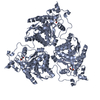
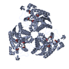
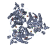
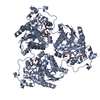
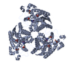
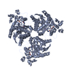
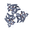
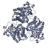
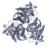
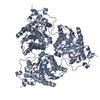
| 構造データ | PDB-4q3p:
Crystal structure of Schistosoma mansoni arginase
手法: X-RAY DIFFRACTION / 解像度: 2.501 Å PDB-4q3q:
Crystal structure of Schistosoma mansoni arginase in complex with inhibitor ABH
手法: X-RAY DIFFRACTION / 解像度: 2.001 Å PDB-4q3r:
Crystal structure of Schistosoma mansoni arginase in complex with inhibitor ABHDP
手法: X-RAY DIFFRACTION / 解像度: 2.169 Å PDB-4q3s:
Crystal structure of Schistosoma mansoni arginase in complex with inhibitor ABHPE
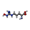
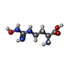
手法: X-RAY DIFFRACTION / 解像度: 2.11 Å PDB-4q3t:
Crystal structure of Schistosoma mansoni arginase in complex with inhibitor NOHA
手法: X-RAY DIFFRACTION / 解像度: 2.142 Å PDB-4q3u:
Crystal structure of Schistosoma mansoni arginase in complex with inhibitor nor-NOHA
手法: X-RAY DIFFRACTION / 解像度: 2.5 Å PDB-4q3v:
Crystal structure of Schistosoma mansoni arginase in complex with inhibitor BEC
手法: X-RAY DIFFRACTION / 解像度: 2.7 Å PDB-4q40:
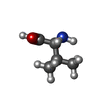
Crystal structure of Schistosoma mansoni arginase in complex with L-valine
手法: X-RAY DIFFRACTION / 解像度: 1.831 Å PDB-4q41:
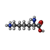
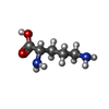
Crystal structure of Schistosoma mansoni arginase in complex with L-lysine
手法: X-RAY DIFFRACTION / 解像度: 2.199 Å PDB-4q42:
Crystal structure of Schistosoma mansoni arginase in complex with L-ornithine
手法: X-RAY DIFFRACTION / 解像度: 2.051 Å |
|---|
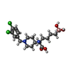
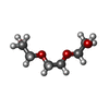
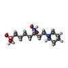
| 化合物 | ChemComp-XA2:
(R)-2-amino-6-borono-2-(1-(3,4-dichlorobenzyl)piperidin-4-yl)hexanoic acid
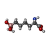
ChemComp-X7A:
[(5R)-5-amino-5-carboxy-7-(piperidin-1-yl)heptyl](trihydroxy)borate(1-)
|
|---|
| 由来 |   schistosoma mansoni (マンソン住血吸虫) schistosoma mansoni (マンソン住血吸虫)
|
|---|
 キーワード キーワード | HYDROLASE / arginase-deacetylase fold / HYDROLASE/HYDROLASE INHIBITOR / HYDROLASE-HYDROLASE INHIBITOR complex |
|---|
 著者
著者 リンク
リンク Biochemistry /
  PubMed:25007099





 キーワード
キーワード ムービー
ムービー コントローラー
 構造ビューア
 万見文献について


