 ムービー
ムービー コントローラー
 構造ビューア
 万見文献について


+検索条件
-Structure paper
| タイトル | Structural model of human endoglin, a transmembrane receptor responsible for hereditary hemorrhagic telangiectasia. |
|---|---|
| ジャーナル・号・ページ | J Mol Biol, Vol. 365, Issue 3, Page 694-705, Year 2007 |
| 掲載日 | 2007年1月19日 |
 著者 著者 | Oscar Llorca / Arturo Trujillo / Francisco J Blanco / Carmelo Bernabeu /  |
| PubMed 要旨 | Endoglin is a type I membrane protein expressed as a disulphide-linked homodimer on human vascular endothelial cells whose haploinsufficiency is responsible for the dominant vascular dysplasia known ...Endoglin is a type I membrane protein expressed as a disulphide-linked homodimer on human vascular endothelial cells whose haploinsufficiency is responsible for the dominant vascular dysplasia known as hereditary hemorrhagic telangiectasia (HHT). Structurally, endoglin belongs to the zona pellucida (ZP) family of proteins that share a ZP domain of approximately 260 amino acid residues at their extracellular region. Endoglin is a component of the TGF-beta receptor complex, interacts with the TGF-beta signalling receptors types I and II, and modulates cellular responses to TGF-beta. Here, we have determined for the first time the three-dimensional structure of the approximately 140 kDa extracellular domain of endoglin at 25 A resolution, using single-particle electron microscopy (EM). This reconstruction provides the general architecture of endoglin, which arranges as a dome made of antiparallel oriented monomers enclosing a cavity at one end. A high-resolution structure of endoglin has also been modelled de novo and found to be consistent with the experimental reconstruction. Each subunit comprises three well-defined domains, two of them corresponding to ZP regions, organised into an open U-shaped monomer. This domain arrangement was found to closely resemble the overall structure derived experimentally and the three modelled de novo domains were tentatively assigned to the domains observed in the EM reconstruction. This molecular model was further tested by tagging endoglin's C terminus with an IgG Fc fragment visible after 3D reconstruction of the labelled protein. Combined, these data provide the structural framework to interpret endoglin's functional domains and mutations found in HHT patients. |
 リンク リンク | J Mol Biol / PubMed:17081563 |
| 手法 | EM (単粒子) |
| 解像度 | 25.0 - 30.0 Å |
| 構造データ | 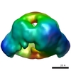 EMDB-1559: 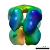 EMDB-1560: |
| 由来 |
|
 Homo sapiens (ヒト)