[English] 日本語
Yorodumi Papers
- Database of articles cited by EMDB/PDB/SASBDB data -

+
Search query

Keywords
Structure methods
Author
Journal
IF

-
Structure paper

TitleThe molecular basis of protein toxin HicA-dependent binding of the protein antitoxin HicB to DNA.
Journal, issue, pagesJ Biol Chem, Vol. 293, Issue 50, Page 19429-19440, Year 2018
Publish dateDec 14, 2018
AuthorsAshley J Winter / Christopher Williams / Michail N Isupov / Hannah Crocker / Mariya Gromova / Philip Marsh / Oliver J Wilkinson / Mark S Dillingham / Nicholas J Harmer / Richard W Titball / Matthew P Crump /
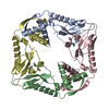
PubMed AbstractToxin-antitoxin (TA) systems are present in many bacteria and play important roles in bacterial growth, physiology, and pathogenicity. Those that are best studied are the type II TA systems, in which ...Toxin-antitoxin (TA) systems are present in many bacteria and play important roles in bacterial growth, physiology, and pathogenicity. Those that are best studied are the type II TA systems, in which both toxins and antitoxins are proteins. The HicAB system is one of the prototypic TA systems, found in many bacterial species. Complex interactions between the protein toxin (HicA), the protein antitoxin (HicB), and the DNA upstream of the encoding genes regulate the activity of this system, but few structural details are available about how HicA destabilizes the HicB-DNA complex. Here, we determined the X-ray structures of HicB and the HicAB complex to 1.8 and 2.5 Å resolution, respectively, and characterized their DNA interactions. This revealed that HicB forms a tetramer and HicA and HicB form a heterooctameric complex that involves structural reorganization of the C-terminal (DNA-binding) region of HicB. Our observations indicated that HicA has a profound impact on binding of HicB to DNA sequences upstream of in a stoichiometric-dependent way. At low ratios of HicA:HicB, there was no effect on DNA binding, but at higher ratios, the affinity for DNA declined cooperatively, driving dissociation of the HicA:HicB:DNA complex. These results reveal the structural mechanisms by which HicA de-represses the HicB-DNA complex.
External linksJ Biol Chem / PubMed:30337369 / PubMed Central
MethodsSAS (X-ray synchrotron) / X-ray diffraction
Resolution1.56 - 2.49 Å
Structure data

SASDD45:
Type II toxin-antitoxin system HicB family antitoxin - HicB protein
Method: SAXS/SANS

SASDD55:
Type II toxin-antitoxin system HicB family antitoxin - HicB protein - bound to addiction module toxin, HicA
Method: SAXS/SANS

PDB-6g1c:
Crystal structure of the N-terminal domain of Burkholderia Pseudomallei antitoxin HicB
Method: X-RAY DIFFRACTION / Resolution: 1.56 Å

PDB-6g1n:
Crystal structure of the Burkholderia Pseudomallei antitoxin HicB
Method: X-RAY DIFFRACTION / Resolution: 1.85 Å

PDB-6g26:
The crystal structure of the Burkholderia pseudomallei HicAB complex
Method: X-RAY DIFFRACTION / Resolution: 2.49 Å

Chemicals

ChemComp-HOH:
WATER

ChemComp-CL:
Unknown entry

ChemComp-GOL:
GLYCEROL

ChemComp-SO4:
SULFATE ION

ChemComp-EDO:
1,2-ETHANEDIOL

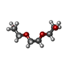
ChemComp-PGE:
TRIETHYLENE GLYCOL

Source
  • burkholderia pseudomallei (bacteria)
  • burkholderia pseudomallei k96243 (bacteria)
KeywordsANTITOXIN / N-terminal domain of the antitoxin HicB which acts as an inhibitor to HicA / The antitoxin HicB which acts as an inhibitor to HicA

+
About Yorodumi Papers

-
News

-
Feb 9, 2022. New format data for meta-information of EMDB entries

New format data for meta-information of EMDB entries

  • Version 3 of the EMDB header file is now the official format.
  • The previous official version 1.9 will be removed from the archive.

Related info.:EMDB header

External links:wwPDB to switch to version 3 of the EMDB data model

-
Aug 12, 2020. Covid-19 info

Covid-19 info

URL: https://pdbj.org/emnavi/covid19.php

New page: Covid-19 featured information page in EM Navigator.

Related info.:Covid-19 info / Mar 5, 2020. Novel coronavirus structure data

+
Mar 5, 2020. Novel coronavirus structure data

Novel coronavirus structure data

Related info.:Yorodumi Speices / Aug 12, 2020. Covid-19 info

External links:COVID-19 featured content - PDBj / Molecule of the Month (242):Coronavirus Proteases

+
Jan 31, 2019. EMDB accession codes are about to change! (news from PDBe EMDB page)

EMDB accession codes are about to change! (news from PDBe EMDB page)

  • The allocation of 4 digits for EMDB accession codes will soon come to an end. Whilst these codes will remain in use, new EMDB accession codes will include an additional digit and will expand incrementally as the available range of codes is exhausted. The current 4-digit format prefixed with “EMD-” (i.e. EMD-XXXX) will advance to a 5-digit format (i.e. EMD-XXXXX), and so on. It is currently estimated that the 4-digit codes will be depleted around Spring 2019, at which point the 5-digit format will come into force.
  • The EM Navigator/Yorodumi systems omit the EMD- prefix.

Related info.:Q: What is EMD? / ID/Accession-code notation in Yorodumi/EM Navigator

External links:EMDB Accession Codes are Changing Soon! / Contact to PDBj

+
Jul 12, 2017. Major update of PDB

Major update of PDB

  • wwPDB released updated PDB data conforming to the new PDBx/mmCIF dictionary.
  • This is a major update changing the version number from 4 to 5, and with Remediation, in which all the entries are updated.
  • In this update, many items about electron microscopy experimental information are reorganized (e.g. em_software).
  • Now, EM Navigator and Yorodumi are based on the updated data.

External links:wwPDB Remediation / Enriched Model Files Conforming to OneDep Data Standards Now Available in the PDB FTP Archive

-
Yorodumi Papers

Database of articles cited by EMDB/PDB/SASBDB data

  • Database of articles cited by EMDB, PDB, and SASBDB entries
  • Using PubMed data

Related info.:EMDB / PDB / SASBDB / Yorodumi / EMN Papers / Changes in new EM Navigator and Yorodumi

Read more