 ムービー
ムービー コントローラー
 構造ビューア
 万見文献について


+検索条件
-Structure paper
| タイトル | Rapid simulation of glycoprotein structures by grafting and steric exclusion of glycan conformer libraries. |
|---|---|
| ジャーナル・号・ページ | Cell, Vol. 187, Issue 5, Page 1296-1311.e26, Year 2024 |
| 掲載日 | 2024年2月29日 |
 著者 著者 | Yu-Xi Tsai / Ning-En Chang / Klaus Reuter / Hao-Ting Chang / Tzu-Jing Yang / Sören von Bülow / Vidhi Sehrawat / Noémie Zerrouki / Matthieu Tuffery / Michael Gecht / Isabell Louise Grothaus / Lucio Colombi Ciacchi / Yong-Sheng Wang / Min-Feng Hsu / Kay-Hooi Khoo / Gerhard Hummer / Shang-Te Danny Hsu / Cyril Hanus / Mateusz Sikora /      |
| PubMed 要旨 | Most membrane proteins are modified by covalent addition of complex sugars through N- and O-glycosylation. Unlike proteins, glycans do not typically adopt specific secondary structures and remain ...Most membrane proteins are modified by covalent addition of complex sugars through N- and O-glycosylation. Unlike proteins, glycans do not typically adopt specific secondary structures and remain very mobile, shielding potentially large fractions of protein surface. High glycan conformational freedom hinders complete structural elucidation of glycoproteins. Computer simulations may be used to model glycosylated proteins but require hundreds of thousands of computing hours on supercomputers, thus limiting routine use. Here, we describe GlycoSHIELD, a reductionist method that can be implemented on personal computers to graft realistic ensembles of glycan conformers onto static protein structures in minutes. Using molecular dynamics simulation, small-angle X-ray scattering, cryoelectron microscopy, and mass spectrometry, we show that this open-access toolkit provides enhanced models of glycoprotein structures. Focusing on N-cadherin, human coronavirus spike proteins, and gamma-aminobutyric acid receptors, we show that GlycoSHIELD can shed light on the impact of glycans on the conformation and activity of complex glycoproteins. |
 リンク リンク | Cell / PubMed:38428397 |
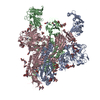
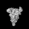
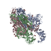
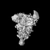
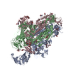
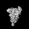
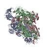
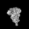
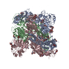
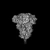
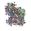
| 手法 | EM (単粒子) |
| 解像度 | 4.1 - 6.87 Å |
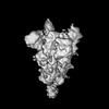
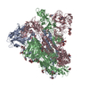
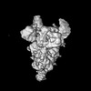
| 構造データ | EMDB-33942, PDB-7ymt: EMDB-33943, PDB-7ymv: EMDB-33944, PDB-7ymw: 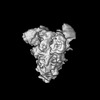 EMDB-33945: Cryo-EM structure of MERS-CoV spike protein, One RBD-up conformation 3 EMDB-33946, PDB-7ymx: EMDB-33947, PDB-7ymy: EMDB-33948, PDB-7ymz: EMDB-33949, PDB-7yn0:  EMDB-38650: Additional map for SARS-CoV-2 Spike D614G variant, one RBD-up conformation 1 (PDB ID: 7EAZ; EMD-31047). Map was generated from heterogeneous refinement with downsampling in CryoSPARC |
| 化合物 |  ChemComp-NAG: |
| 由来 |
|
 キーワード キーワード | VIRAL PROTEIN / MERS-CoV / Spike / Glycoprotein |
 human betacoronavirus 2c emc/2012 (ウイルス)
 Severe acute respiratory syndrome coronavirus 2 (ウイルス)