 ムービー
ムービー コントローラー
 構造ビューア
 万見文献について


+検索条件
-Structure paper
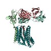
| タイトル | The tertiary structure of the human Xkr8-Basigin complex that scrambles phospholipids at plasma membranes. |
|---|---|
| ジャーナル・号・ページ | Nat Struct Mol Biol, Vol. 28, Issue 10, Page 825-834, Year 2021 |
| 掲載日 | 2021年10月8日 |
 著者 著者 | Takaharu Sakuragi / Ryuta Kanai / Akihisa Tsutsumi / Hirotaka Narita / Eriko Onishi / Kohei Nishino / Takuya Miyazaki / Takeshi Baba / Hidetaka Kosako / Atsushi Nakagawa / Masahide Kikkawa / Chikashi Toyoshima / Shigekazu Nagata /  |
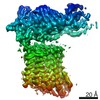
| PubMed 要旨 | Xkr8-Basigin is a plasma membrane phospholipid scramblase activated by kinases or caspases. We combined cryo-EM and X-ray crystallography to investigate its structure at an overall resolution of 3. ...Xkr8-Basigin is a plasma membrane phospholipid scramblase activated by kinases or caspases. We combined cryo-EM and X-ray crystallography to investigate its structure at an overall resolution of 3.8 Å. Its membrane-spanning region carrying 22 charged amino acids adopts a cuboid-like structure stabilized by salt bridges between hydrophilic residues in transmembrane helices. Phosphatidylcholine binding was observed in a hydrophobic cleft on the surface exposed to the outer leaflet of the plasma membrane. Six charged residues placed from top to bottom inside the molecule were essential for scrambling phospholipids in inward and outward directions, apparently providing a pathway for their translocation. A tryptophan residue was present between the head group of phosphatidylcholine and the extracellular end of the path. Its mutation to alanine made the Xkr8-Basigin complex constitutively active, indicating that it plays a vital role in regulating its scramblase activity. The structure of Xkr8-Basigin provides insights into the molecular mechanisms underlying phospholipid scrambling. |
 リンク リンク | Nat Struct Mol Biol / PubMed:34625749 / PubMed Central |
| 手法 | EM (単粒子) / X線回折 |
| 解像度 | 1.123 - 3.8 Å |
| 構造データ | EMDB-30636, PDB-7dce: 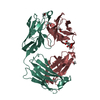 PDB-7d9z: 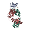 PDB-7daa: |
| 化合物 |  ChemComp-EDO:  ChemComp-FLC:  ChemComp-HOH:  ChemComp-CD: 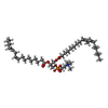 ChemComp-DLP: |
| 由来 |
|
 キーワード キーワード | IMMUNE SYSTEM / Fab / antibody / basigin / CHAPERONE / complex / TRANSPORT PROTEIN / XKR8 / scramblase / phospholipid |
 homo sapiens (ヒト)
