 Movie
Movie Controller
 Structure viewers
 About Yorodumi Papers


+Search query
-Structure paper
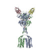
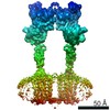
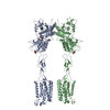
| Title | Structural insights into the activation of metabotropic glutamate receptors. |
|---|---|
| Journal, issue, pages | Nature, Vol. 566, Issue 7742, Page 79-84, Year 2019 |
| Publish date | Jan 23, 2019 |
 Authors Authors | Antoine Koehl / Hongli Hu / Dan Feng / Bingfa Sun / Yan Zhang / Michael J Robertson / Matthew Chu / Tong Sun Kobilka / Toon Laeremans / Jan Steyaert / Jeffrey Tarrasch / Somnath Dutta / Rasmus Fonseca / William I Weis / Jesper M Mathiesen / Georgios Skiniotis / Brian K Kobilka /     |
| PubMed Abstract | Metabotropic glutamate receptors are family C G-protein-coupled receptors. They form obligate dimers and possess extracellular ligand-binding Venus flytrap domains, which are linked by cysteine-rich ...Metabotropic glutamate receptors are family C G-protein-coupled receptors. They form obligate dimers and possess extracellular ligand-binding Venus flytrap domains, which are linked by cysteine-rich domains to their 7-transmembrane domains. Spectroscopic studies show that signalling is a dynamic process, in which large-scale conformational changes underlie the transmission of signals from the extracellular Venus flytraps to the G protein-coupling domains-the 7-transmembrane domains-in the membrane. Here, using a combination of X-ray crystallography, cryo-electron microscopy and signalling studies, we present a structural framework for the activation mechanism of metabotropic glutamate receptor subtype 5. Our results show that agonist binding at the Venus flytraps leads to a compaction of the intersubunit dimer interface, thereby bringing the cysteine-rich domains into close proximity. Interactions between the cysteine-rich domains and the second extracellular loops of the receptor enable the rigid-body repositioning of the 7-transmembrane domains, which come into contact with each other to initiate signalling. |
 External links External links | Nature / PubMed:30675062 / PubMed Central |
| Methods | EM (single particle) / X-ray diffraction |
| Resolution | 3.262 - 7.9 Å |
| Structure data | EMDB-0345, PDB-6n51: EMDB-0346, PDB-6n52: 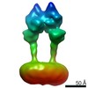 EMDB-0347: 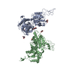 PDB-6n4x: 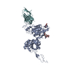 PDB-6n4y: 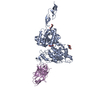 PDB-6n50: |
| Chemicals |  ChemComp-NAG:  ChemComp-MG:  ChemComp-QUS: |
| Source |
|
 Keywords Keywords | MEMBRANE PROTEIN / Cell Surface Receptor / Cell Surface Receptor Nanobody / SIGNALING PROTEIN |

 homo sapiens (human)
