 ムービー
ムービー コントローラー
 構造ビューア
 万見文献について


+検索条件
-Structure paper
| タイトル | Small-angle X-ray scattering study of the kinetics of light-dark transition in a LOV protein. |
|---|---|
| ジャーナル・号・ページ | PLoS One, Vol. 13, Issue 7, Page e0200746, Year 2018 |
| 掲載日 | 2018年7月16日 |
 著者 著者 | Katrin Röllen / Joachim Granzin / Renu Batra-Safferling / Andreas Maximilian Stadler /  |
| PubMed 要旨 | Light, oxygen, voltage (LOV) photoreceptors consist of conserved photo-responsive domains in bacteria, archaea, plants and fungi, and detect blue-light via a flavin cofactor. We investigated the blue- ...Light, oxygen, voltage (LOV) photoreceptors consist of conserved photo-responsive domains in bacteria, archaea, plants and fungi, and detect blue-light via a flavin cofactor. We investigated the blue-light induced conformational transition of the dimeric photoreceptor PpSB1-LOV-R66I from Pseudomonas putida in solution by using small-angle X-ray scattering (SAXS). SAXS experiments of the fully populated light- and dark-states under steady-state conditions revealed significant structural differences between the two states that are in agreement with the known structures determined by crystallography. We followed the transition from the light- to the dark-state by using SAXS measurements in real-time. A two-state model based on the light- and dark-state conformations could describe the measured time-course SAXS data with a relaxation time τREC of ~ 34 to 35 min being larger than the recovery time found with UV/vis spectroscopy. Unlike the flavin chromophore-based UV/vis method that is sensitive to the local chromophore environment in flavoproteins, SAXS-based assay depends on protein conformational changes and provides with an alternative to measure the recovery kinetics. |
 リンク リンク | PLoS One / PubMed:30011332 / PubMed Central |
| 手法 | SAS (X-ray synchrotron) / X線回折 |
| 解像度 | 2.04 Å |
| 構造データ |  SASDDE6:  SASDDG6: 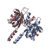 PDB-6gg9: |
| 化合物 |  ChemComp-FMN:  ChemComp-HOH: |
| 由来 |
|
 キーワード キーワード | SIGNALING PROTEIN / blue light photoreceptor / Sensory box protein |
 Pseudomonas putida (strain atcc 47054 / dsm 6125 / ncimb 11950 / kt2440) (バクテリア)