| タイトル | "Newton's cradle" proton relay with amide-imidic acid tautomerization in inverting cellulase visualized by neutron crystallography. |
|---|
| ジャーナル・号・ページ | Sci Adv, Vol. 1, Page e1500263-e1500263, Year 2015 |
|---|
| 掲載日 | 2014年12月22日 (構造データの登録日) |
|---|
 著者 著者 | Nakamura, A. / Ishida, T. / Kusaka, K. / Yamada, T. / Fushinobu, S. / Tanaka, I. / Kaneko, S. / Ohta, K. / Tanaka, H. / Inaka, K. ...Nakamura, A. / Ishida, T. / Kusaka, K. / Yamada, T. / Fushinobu, S. / Tanaka, I. / Kaneko, S. / Ohta, K. / Tanaka, H. / Inaka, K. / Higuchi, Y. / Niimura, N. / Samejima, M. / Igarashi, K. |
|---|
 リンク リンク |  Sci Adv / Sci Adv /  PubMed:26601228 PubMed:26601228 |
|---|
| 手法 | X線回折 / 中性子線回折 |
|---|
| 解像度 | 0.64 - 1.6 Å |
|---|
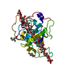
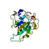
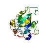
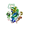
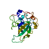
| 構造データ | PDB-3x2g:
X-ray structure of PcCel45A N92D apo form at 100K
手法: X-RAY DIFFRACTION / 解像度: 1 Å PDB-3x2h:
X-ray structure of PcCel45A N92D with cellopentaose at 95K.
手法: X-RAY DIFFRACTION / 解像度: 0.99 Å PDB-3x2i:
X-ray structure of PcCel45A N92D apo form at 298K.
手法: X-RAY DIFFRACTION / 解像度: 1.6 Å PDB-3x2j:
X-ray structure of PcCel45A D114N apo form at 95K.
手法: X-RAY DIFFRACTION / 解像度: 1.301 Å PDB-3x2k:
X-ray structure of PcCel45A D114N with cellopentaose at 95K.
手法: X-RAY DIFFRACTION / 解像度: 1.182 Å PDB-3x2l:
X-ray structure of PcCel45A apo form at 95K.
手法: X-RAY DIFFRACTION / 解像度: 0.83 Å PDB-3x2m:
X-ray structure of PcCel45A with cellopentaose at 0.64 angstrom resolution.
手法: X-RAY DIFFRACTION / 解像度: 0.64 Å PDB-3x2n:
Proton relay pathway in inverting cellulase
手法: X-RAY DIFFRACTION / 解像度: 1.2 Å PDB-3x2o:
Neutron and X-ray joint refined structure of PcCel45A apo form at 298K.
手法: NEUTRON DIFFRACTION / X-RAY DIFFRACTION / 解像度: 1.5 Å PDB-3x2p:
Neutron and X-ray joint refined structure of PcCel45A with cellopentaose at 298K.
手法: NEUTRON DIFFRACTION / X-RAY DIFFRACTION / 解像度: 1.518 Å PDB-4zm7:
PcCel45A N105D mutatnt at cryo condition
手法: X-RAY DIFFRACTION / 解像度: 0.701 Å |
|---|
| 化合物 | |
|---|
| 由来 |  phanerochaete chrysosporium (菌類) phanerochaete chrysosporium (菌類)
|
|---|
 キーワード キーワード | HYDROLASE / Cellulase Endo-glucanase |
|---|
 著者
著者 リンク
リンク Sci Adv /
  PubMed:26601228





 キーワード
キーワード ムービー
ムービー コントローラー
 構造ビューア
 万見文献について


 phanerochaete chrysosporium (菌類)