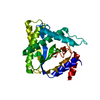
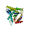
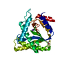
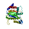
PDB-3wrn: Minute virus of mice non-structural protein-1N-terminal nuclease domain reveals a unique Zn2+ coordination in the active site pocket and shows a novel mode of DNA recognition at the origin of replication 手法: X-RAY DIFFRACTION / 解像度: 1.52 Å
PDB-3wro: Minute virus of mice non-structural protein-1N-terminal nuclease domain reveals a unique Zn2+ coordination in the active site pocket and shows a novel mode of DNA recognition at the origin of replication 手法: X-RAY DIFFRACTION / 解像度: 1.48 Å
PDB-3wrq: Minute virus of mice non-structural protein-1N-terminal nuclease domain reveals a unique Zn2+ coordination in the active site pocket and shows a novel mode of DNA recognition at the origin of replication 手法: X-RAY DIFFRACTION / 解像度: 1.53 Å
PDB-3wrr: Minute virus of mice non-structural protein-1N-terminal nuclease domain reveals a unique Zn2+ coordination in the active site pocket and shows a novel mode of DNA recognition at the origin of replication 手法: X-RAY DIFFRACTION / 解像度: 1.62 Å
PDB-3wrs: Minute virus of mice non-structural protein-1N-terminal nuclease domain reveals a unique Zn2+ coordination in the active site pocket and shows a novel mode of DNA recognition at the origin of replication 手法: X-RAY DIFFRACTION / 解像度: 1.58 Å
PDB-4pp4: Minute virus of mice non-structural protein-1N-terminal nuclease domain reveals a unique Zn2+ coordination in the active site pocket and shows a novel mode of DNA recognition at the origin of replication 手法: X-RAY DIFFRACTION / 解像度: 1.45 Å
PDB-4r94: Structure of the nickase domain of NS1 from MVM complexed with magnesium 手法: X-RAY DIFFRACTION / 解像度: 1.668 Å
REPLICATION / Nuclease activity / Single and double stranded DNA binding / nicking protein / Single and double stranded binding / Single and double stranded DNA binding and nicking protein / single/double strand DNA binding / nickase domain / DNA binding / magnesium / nickase
 ムービー
ムービー コントローラー
 構造ビューア
 万見文献について


 著者
著者 リンク
リンク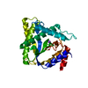


 キーワード
キーワード murine minute virus (マウス微小ウイルス)