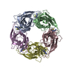
Studies on an acetylcholine binding protein identify a basic residue in loop G on the beta 1 strand as a new structural determinant of neonicotinoid actions
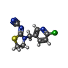
PDB-3wth: Crystal Structure of Lymnaea stagnalis Acetylcholine-Binding Protein Q55R Mutant Complexed with Imidacloprid 手法: X-RAY DIFFRACTION / 解像度: 2.54 Å
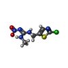
PDB-3wti: Crystal Structure of Lymnaea stagnalis Acetylcholine-Binding Protein Q55R Mutant Complexed with Clothianidin 手法: X-RAY DIFFRACTION / 解像度: 2.68 Å
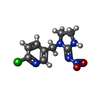
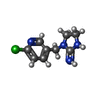
PDB-3wtj: Crystal Structure of Lymnaea stagnalis Acetylcholine Binding Protein Complexed with Thiacloprid 手法: X-RAY DIFFRACTION / 解像度: 2.24 Å
PDB-3wtk: Crystal Structure of Lymnaea stagnalis Acetylcholine-Binding Protein Q55R Mutant Complexed with Thiacloprid 手法: X-RAY DIFFRACTION / 解像度: 2.69 Å
PDB-3wtl: Crystal Structure of Lymnaea stagnalis Acetylcholine Binding Protein Complexed with Nitromethylene Analogue of Imidacloprid 手法: X-RAY DIFFRACTION / 解像度: 2.3 Å
PDB-3wtm: Crystal Structure of Lymnaea stagnalis Acetylcholine-Binding Protein Q55R Mutant Complexed with Nitromethylene Analogue of Imidacloprid 手法: X-RAY DIFFRACTION / 解像度: 2.48 Å
PDB-3wtn: Crystal Structure of Lymnaea stagnalis Acetylcholine Binding Protein Complexed with Desnitro-imidacloprid 手法: X-RAY DIFFRACTION / 解像度: 2.09 Å
PDB-3wto: Crystal Structure of Lymnaea stagnalis Acetylcholine-Binding Protein Q55R Mutant Complexed with Desnitro-imidacloprid 手法: X-RAY DIFFRACTION / 解像度: 2.25 Å
 ムービー
ムービー コントローラー
 構造ビューア
 万見文献について


 著者
著者 リンク
リンク


 キーワード
キーワード
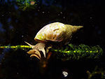 lymnaea stagnalis (無脊椎動物)