ムービー
ムービー コントローラー
 構造ビューア
 万見文献について


+検索条件
-Structure paper
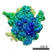
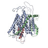
| タイトル | Visualization of a polytopic membrane protein during SecY-mediated membrane insertion. |
|---|---|
| ジャーナル・号・ページ | Nat Commun, Vol. 5, Page 4103, Year 2014 |
| 掲載日 | 2014年6月10日 |
 著者 著者 | Lukas Bischoff / Stephan Wickles / Otto Berninghausen / Eli O van der Sluis / Roland Beckmann /  |
| PubMed 要旨 | The biogenesis of polytopic membrane proteins occurs co-translationally on ribosomes that are tightly bound to a membrane-embedded protein-conducting channel: the Sec-complex. The path that is ...The biogenesis of polytopic membrane proteins occurs co-translationally on ribosomes that are tightly bound to a membrane-embedded protein-conducting channel: the Sec-complex. The path that is followed by nascent proteins inside the ribosome and the Sec-complex is relatively well established; however, it is not clear what the fate of the N-terminal transmembrane domains (TMDs) of polytopic membrane proteins is when the C-terminal TMDs domains are not yet synthesized. Here, we present the sub-nanometer cryo-electron microscopy structure of an in vivo generated ribosome-SecY complex that carries a membrane insertion intermediate of proteorhodopsin (PR). The structure reveals a pre-opened Sec-complex and the first two TMDs of PR already outside the SecY complex directly in front of its proposed lateral gate. Thus, our structure is in agreement with positioning of N-terminal TMDs at the periphery of SecY, and in addition, it provides clues for the molecular mechanism underlying membrane protein topogenesis. |
 リンク リンク | Nat Commun / PubMed:24912953 |
| 手法 | EM (単粒子) |
| 解像度 | 7.28 - 8 Å |
| 構造データ | |
| 由来 |
|
 キーワード キーワード | TRANSLATION / RIBOSOME / MEMBRANE PROTEIN / TRANSLOCON |