 Movie
Movie Controller

[English] 日本語
 Yorodumi
- PDB-5abb: Visualization of a polytopic membrane protein during SecY-mediate... -
+ Open data

- Basic information
| Entry | Database: PDB / ID: 5abb | ||||||
|---|---|---|---|---|---|---|---|
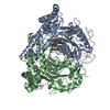
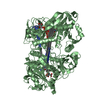
| Title | Visualization of a polytopic membrane protein during SecY-mediated membrane insertion | ||||||
 Components Components |
| ||||||
 Keywords Keywords | TRANSLATION / RIBOSOME / MEMBRANE PROTEIN / TRANSLOCON | ||||||
| Function / homology |  Function and homology information Function and homology informationlight-activated monoatomic ion channel activity / cell envelope Sec protein transport complex / protein transport by the Sec complex / intracellular protein transmembrane transport / SRP-dependent cotranslational protein targeting to membrane, translocation / signal sequence receptor activity / Secretion of toxins / photoreceptor activity / transmembrane protein transporter activity / protein secretion ...light-activated monoatomic ion channel activity / cell envelope Sec protein transport complex / protein transport by the Sec complex / intracellular protein transmembrane transport / SRP-dependent cotranslational protein targeting to membrane, translocation / signal sequence receptor activity / Secretion of toxins / photoreceptor activity / transmembrane protein transporter activity / protein secretion / phototransduction / protein targeting / proton transmembrane transport / intracellular protein transport / membrane / plasma membrane Similarity search - Function | ||||||
| Biological species |  | ||||||
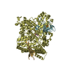
| Method | ELECTRON MICROSCOPY / single particle reconstruction / cryo EM / Resolution: 8 Å | ||||||
 Authors Authors | Bischoff, L. / Wickles, S. / Berninghausen, O. / vanderSluis, E. / Beckmann, R. | ||||||
 Citation Citation | Journal: Nat Commun / Year: 2014 Title: Visualization of a polytopic membrane protein during SecY-mediated membrane insertion. Authors: Lukas Bischoff / Stephan Wickles / Otto Berninghausen / Eli O van der Sluis / Roland Beckmann /  Abstract: The biogenesis of polytopic membrane proteins occurs co-translationally on ribosomes that are tightly bound to a membrane-embedded protein-conducting channel: the Sec-complex. The path that is ...The biogenesis of polytopic membrane proteins occurs co-translationally on ribosomes that are tightly bound to a membrane-embedded protein-conducting channel: the Sec-complex. The path that is followed by nascent proteins inside the ribosome and the Sec-complex is relatively well established; however, it is not clear what the fate of the N-terminal transmembrane domains (TMDs) of polytopic membrane proteins is when the C-terminal TMDs domains are not yet synthesized. Here, we present the sub-nanometer cryo-electron microscopy structure of an in vivo generated ribosome-SecY complex that carries a membrane insertion intermediate of proteorhodopsin (PR). The structure reveals a pre-opened Sec-complex and the first two TMDs of PR already outside the SecY complex directly in front of its proposed lateral gate. Thus, our structure is in agreement with positioning of N-terminal TMDs at the periphery of SecY, and in addition, it provides clues for the molecular mechanism underlying membrane protein topogenesis. | ||||||
| History |
|
- Structure visualization
| Movie |
Movie viewer |
|---|---|
| Structure viewer | Molecule: MolmilJmol/JSmol |
- Downloads & links
-Download
| PDBx/mmCIF format | 5abb.cif.gz | 90.6 KB | Display | PDBx/mmCIF format |
|---|---|---|---|---|
| PDB format | pdb5abb.ent.gz | 54.9 KB | Display | PDB format |
| PDBx/mmJSON format | 5abb.json.gz | Tree view | PDBx/mmJSON format | |
| Others |  Other downloads Other downloads |
-Validation report
| Arichive directory | https://data.pdbj.org/pub/pdb/validation_reports/ab/5abbftp://data.pdbj.org/pub/pdb/validation_reports/ab/5abb | HTTPS FTP |
|---|
-Related structure data
| Related structure data | 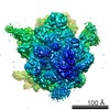 2446MC M: map data used to model this data C: citing same article ( |
|---|---|
| Similar structure data |


-Links
 PDBj
PDBj


- Assembly
| Deposited unit | 
|
|---|---|
| 1 |
|
-Components
| #1: Protein | Mass: 48553.375 Da / Num. of mol.: 1 / Source method: isolated from a natural source / Source: (natural) |
|---|---|
| #2: Protein | Mass: 12623.296 Da / Num. of mol.: 1 / Fragment: UNP RESIDUES 12-127 / Source method: isolated from a natural source / Source: (natural) |
| #3: Protein | Mass: 7634.684 Da / Num. of mol.: 1 / Fragment: UNP RESIDUES 19-83 / Source method: isolated from a natural source / Source: (natural) |
-Experimental details
-Experiment
| Experiment | Method: ELECTRON MICROSCOPY |
|---|---|
| EM experiment | Aggregation state: PARTICLE / 3D reconstruction method: single particle reconstruction |
- Sample preparation
| Component | Name: TnaC stalled E.coli ribosome in complex with SecYE / Type: RIBOSOME |
|---|---|
| Buffer solution | Name: 20 MM TRIS 150 MM NH4CL 10 MM MGCL2 0.05%DDM 125 MM SUCROSE pH: 7.5 Details: 20 MM TRIS 150 MM NH4CL 10 MM MGCL2 0.05%DDM 125 MM SUCROSE |
| Specimen | Embedding applied: NO / Shadowing applied: NO / Staining applied: NO / Vitrification applied: YES |
| Specimen support | Details: HOLEY CARBON |
| Vitrification | Instrument: FEI VITROBOT MARK IV / Cryogen name: ETHANE |
- Electron microscopy imaging
| Experimental equipment |  Model: Titan Krios / Image courtesy: FEI Company |
|---|---|
| Microscopy | Model: FEI TITAN KRIOS / Date: May 21, 2012 |
| Electron gun | Electron source:  FIELD EMISSION GUN / Accelerating voltage: 200 kV / Illumination mode: SPOT SCAN FIELD EMISSION GUN / Accelerating voltage: 200 kV / Illumination mode: SPOT SCAN |
| Electron lens | Mode: BRIGHT FIELD / Nominal magnification: 148721 X / Nominal defocus max: 3500 nm / Nominal defocus min: 1000 nm |
| Image recording | Electron dose: 20 e/Å2 / Film or detector model: TVIPS TEMCAM-F416 (4k x 4k) |
| Radiation wavelength | Relative weight: 1 |
- Processing
| EM software |
| ||||||||||||
|---|---|---|---|---|---|---|---|---|---|---|---|---|---|
| Symmetry | Point symmetry: C1 (asymmetric) | ||||||||||||
| 3D reconstruction | Method: REAL / Resolution: 8 Å / Num. of particles: 47471 / Symmetry type: POINT | ||||||||||||
| Refinement | Highest resolution: 8 Å | ||||||||||||
| Refinement step | Cycle: LAST / Highest resolution: 8 Å
|
