 ムービー
ムービー コントローラー
 構造ビューア
 万見文献について


+検索条件
-Structure paper
| タイトル | Immuno- and constitutive proteasome crystal structures reveal differences in substrate and inhibitor specificity. |
|---|---|
| ジャーナル・号・ページ | Cell(Cambridge,Mass. ), Vol. 148, Page 727-738, Year 2012 |
| 掲載日 | 2011年11月15日 (構造データの登録日) |
 著者 著者 | Huber, E.M. / Basler, M. / Schwab, R. / Heinemeyer, W. / Kirk, C.J. / Groettrup, M. / Groll, M. |
 リンク リンク | Cell(Cambridge,Mass. ) / PubMed:22341445 |
| 手法 | X線回折 |
| 解像度 | 2.7 - 3.4 Å |
| 構造データ | 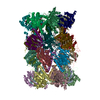 PDB-3un4: 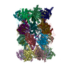 PDB-3un8: 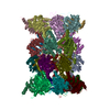 PDB-3unb:  PDB-3une:  PDB-3unf: 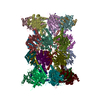 PDB-3unh: |
| 化合物 | 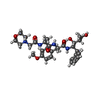 ChemComp-04C:  ChemComp-HOH: 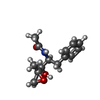 ChemComp-049:  ChemComp-CL:  ChemComp-K:  ChemComp-IOD: |
| 由来 |
|
 キーワード キーワード | HYDROLASE/HYDROLASE INHIBITOR / Proteasome / antigen presentation / drug development / protein degradation / HYDROLASE -HYDROLASE-INHIBITOR complex / HYDROLASE-HYDROLASE INHIBITOR complex / HYDROLASE/HYDROLASE INHIBTIOR / HYDROLASE-HYDROLASE INHIBTIOR complex / 20S proteasome comprises 28 subunits; each subunit adopts the fold of an antiparallel beta-sheet flanked by helices / Protease / Regulatory complexes / Covalent binding of PR-957 to all active sites / HYDROLASE / 20S proteasome comprises 28 subunits / Cytosol |


