 Movie
Movie Controller
 Structure viewers
 About EMN search


-Search query
-Search result
Showing 1 - 50 of 2,815 items for (author: zhu & r)

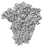
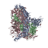
EMDB-45078: 
E.coli GroEL + PBZ1587 inhibitor

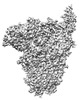
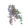
EMDB-45079:
E.coli GroEL apoenzyme
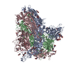
EMDB-45080:
E.Faecium GroEL

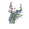
EMDB-45098:
E.coli GroEL + PBZ1587 inhibitor C1 reconstruction
PDB-9c0b:
E.coli GroEL + PBZ1587 inhibitor
PDB-9c0c:
E.coli GroEL apoenzyme
PDB-9c0d:
E.Faecium GroEL
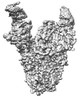
EMDB-39623:
Cryo-EM structure of the retatrutide-bound human GCGR-Gs complex
PDB-8yw5:
Cryo-EM structure of the retatrutide-bound human GCGR-Gs complex
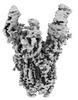
EMDB-60062:
cryo-EM structure of GPR4-Gs complex at pH 8.5
PDB-8zfj:
cryo-EM structure of GPR4-Gs complex at pH 8.5
EMDB-32979:
Cryo-EM structure of Coxsackievirus B1 A-particle in complex with nAb 8A10 (CVB1-A:8A10)
PDB-7x35:
Cryo-EM structure of Coxsackievirus B1 A-particle in complex with nAb 8A10 (CVB1-A:8A10)
EMDB-39025:
Structure of HCoV-HKU1A spike in the functionally anchored-3up conformation with 3TMPRSS2
EMDB-39026:
Local structure of HCoV-HKU1A spike in complex with TMPRSS2 and glycan
EMDB-39036:
Structure of HCoV-HKU1C spike in the functionally anchored-1up conformation with 1TMPRSS2
EMDB-39037:
Structure of HCoV-HKU1C spike in the functionally anchored-2up conformation with 2TMPRSS2
EMDB-39038:
Structure of HCoV-HKU1C spike in the functionally anchored-3up conformation with 2TMPRSS2
EMDB-39039:
Structure of HCoV-HKU1C spike in the functionally anchored-3up conformation with 3TMPRSS2
EMDB-39040:
Local structure of HCoV-HKU1C spike in complex with TMPRSS2 and glycan
EMDB-39041:
Structure of HCoV-HKU1C spike in the inactive-closed conformation
EMDB-39042:
Structure of HCoV-HKU1C spike in the inactive-1up conformation
EMDB-39043:
Structure of HCoV-HKU1C spike in the inactive-2up conformation
EMDB-39044:
Structure of HCoV-HKU1C spike in the glycan-activated-closed conformation
EMDB-39045:
Structure of HCoV-HKU1C spike in the glycan-activated-1up conformation
EMDB-39046:
Structure of HCoV-HKU1C spike in the glycan-activated-2up conformation
EMDB-39047:
Structure of HCoV-HKU1C spike in the glycan-activated-3up conformation
EMDB-39048:
Local structure of HCoV-HKU1C spike in complex with glycan
PDB-8y7x:
Structure of HCoV-HKU1A spike in the functionally anchored-3up conformation with 3TMPRSS2
PDB-8y7y:
Local structure of HCoV-HKU1A spike in complex with TMPRSS2 and glycan
PDB-8y87:
Structure of HCoV-HKU1C spike in the functionally anchored-1up conformation with 1TMPRSS2
PDB-8y88:
Structure of HCoV-HKU1C spike in the functionally anchored-2up conformation with 2TMPRSS2
PDB-8y89:
Structure of HCoV-HKU1C spike in the functionally anchored-3up conformation with 2TMPRSS2
PDB-8y8a:
Structure of HCoV-HKU1C spike in the functionally anchored-3up conformation with 3TMPRSS2
PDB-8y8b:
Local structure of HCoV-HKU1C spike in complex with TMPRSS2 and glycan
PDB-8y8c:
Structure of HCoV-HKU1C spike in the inactive-closed conformation

PDB-8y8d:
Structure of HCoV-HKU1C spike in the inactive-1up conformation
PDB-8y8e:
Structure of HCoV-HKU1C spike in the inactive-2up conformation
PDB-8y8f:
Structure of HCoV-HKU1C spike in the glycan-activated-closed conformation

PDB-8y8g:
Structure of HCoV-HKU1C spike in the glycan-activated-1up conformation
PDB-8y8h:
Structure of HCoV-HKU1C spike in the glycan-activated-2up conformation
PDB-8y8i:
Structure of HCoV-HKU1C spike in the glycan-activated-3up conformation
PDB-8y8j:
Local structure of HCoV-HKU1C spike in complex with glycan

EMDB-19129:
A DNA Robotic Switch with Regulated Autonomous Display of Cytotoxic Ligand Nanopatterns
EMDB-36782:
SID1 transmembrane family member 2
EMDB-36783:
SID1 transmembrane family member 2

EMDB-36784:
SID1 transmembrane family member 2
EMDB-36785:
SID1 transmembrane family member 1
EMDB-36791:
SID1 transmembrane family member 1

EMDB-36792:
SID1 transmembrane family member 1
Pages:
 wwPDB to switch to version 3 of the EMDB data model
