 Movie
Movie Controller
 Structure viewers
 About EMN search


-Search query
-Search result
Showing 1 - 50 of 67 items for (author: gallagher-jones & m)
PDB-7n2d: 
MicroED structure of human zinc finger protein 292 segment (534-542) phased by ARCIMBOLDO-BORGES
Method: electron crystallography / : Richards LS, Flores MD, Zee CT, Glynn C, Gallagher-Jones M, Sawaya MR
PDB-7n2e:
MicroED structure of human CPEB3 segment (154-161) straight polymorph
Method: electron crystallography / : Flores MD, Richards LS, Zee CT, Glynn C, Gallagher-Jones M, Sawaya MR
PDB-7n2f:
MicroED structure of human CPEB3 segment (154-161) straight polymorph phased by ARCIMBOLDO-BORGES
Method: electron crystallography / : Flores MD, Richards LS, Zee CT, Glynn C, Gallagher-Jones M, Sawaya MR
PDB-7n2g:
MicroED structure of human CPEB3 segment(154-161) kinked polymorph phased by ARCIMBOLDO-BORGES
Method: electron crystallography / : Flores MD, Richards LS, Zee CT, Glynn C, Gallagher-Jones M, Sawaya MR
PDB-7n2i:
MicroED structure of human LECT2 (45-53) phased by ARCIMBOLDO-BORGES
Method: electron crystallography / : Richards LS, Flores MD, Zee CT, Glynn C, Gallagher-Jones M, Sawaya MR

PDB-7n2j:
MicroED structure of a mutant mammalian prion segment phased by ARCIMBOLDO-BORGES
Method: electron crystallography / : Richards LS, Flores MD, Zee CT, Glynn C, Gallagher-Jones M, Sawaya MR
PDB-7n2k:
MicroED structure of sequence variant of repeat segment of the yeast prion New1p phased by ARCIMBOLDO-BORGES
Method: electron crystallography / : Flores MD, Richards LS, Zee CT, Glynn C, Gallagher-Jones M, Sawaya MR
PDB-7n2l:
MicroED structure of a mutant mammalian prion segment
Method: electron crystallography / : Flores MD, Richards LS, Zee CT, Glynn C, Gallagher-Jones M, Sawaya MR

PDB-6uop:
OsPYL/RCAR5 (24 - 29) solved by nanobeam diffraction tomography
Method: electron crystallography / : Gallagher-Jones M, Richards LS, Lee S, Rodriguez JA

PDB-6uoq:
OsPYL/RCAR5 residues 24-29 solved from electron diffraction stills
Method: electron crystallography / : Gallagher-Jones M, Richards LS, Lee S, Rodriguez JA

PDB-6uor:
MicroED structure of OsPYL/RCAR5 (24-29) at 3 e-/A^2
Method: electron crystallography / : Gallagher-Jones M, Richards LS, Lee S, Rodriguez JA

PDB-6uos:
MicroED structure of OsPYL/RCAR5 (24-29) at 6 e-/A^2
Method: electron crystallography / : Gallagher-Jones M, Richards LS, Lee S, Rodriguez JA

PDB-6uou:
MicroED structure of OsPYL/RCAR5 (24-29) at 9 e-/A^2
Method: electron crystallography / : Gallagher-Jones M, Richards LS, Lee S, Rodriguez JA

PDB-6uow:
MicroED structure of OsPYL/RCAR5 (24-29) at 12 e-/A^2
Method: electron crystallography / : Gallagher-Jones M, Richards LS, Lee S, Rodriguez JA

EMDB-20900:
human prion protein fibril, M129 variant
Method: helical / : Glynn C, Sawaya MR

PDB-6uur:
Human prion protein fibril, M129 variant
Method: helical / : Glynn C, Sawaya MR, Ge P, Zhou ZH, Rodriguez JA
PDB-6m9i:
L-GSTSTA from degenerate octameric repeats in InaZ, residues 707-712
Method: electron crystallography / : Zee C, Glynn C, Gallagher-Jones M, Miao J, Santiago CG, Cascio D, Gonen T, Sawaya MR, Rodriguez JA

PDB-6m9j:
Racemic-GSTSTA from degenerate octameric repeats in InaZ, residues 707-712
Method: electron crystallography / : Zee C, Glynn C, Gallagher-Jones M, Miao J, Santiago CG, Cascio D, Gonen T, Sawaya MR, Rodriguez JA
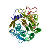
EMDB-7490:
1.71 A MicroED structure of proteinase K at 0.86 e- / A^2
Method: electron crystallography / : Hattne J, Shi D
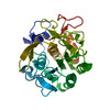
EMDB-7491:
2.00 A MicroED structure of proteinase K at 2.6 e- / A^2
Method: electron crystallography / : Hattne J, Shi D
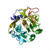
EMDB-7492:
2.20 A MicroED structure of proteinase K at 4.3 e- / A^2
Method: electron crystallography / : Hattne J, Shi D
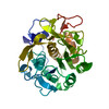
EMDB-7493:
2.80 A MicroED structure of proteinase K at 6.0 e- / A^2
Method: electron crystallography / : Hattne J, Shi D
EMDB-7494:
3.20 A MicroED structure of proteinase K at 7.8 e- / A^2
Method: electron crystallography / : Hattne J, Shi D
EMDB-7495:
1.01 A MicroED structure of GSNQNNF at 0.27 e- / A^2
Method: electron crystallography / : Hattne J, Shi D
EMDB-7496:
1.01 A MicroED structure of GSNQNNF at 0.81 e- / A^2
Method: electron crystallography / : Hattne J, Shi D
EMDB-7497:
1.01 A MicroED structure of GSNQNNF at 1.3 e- / A^2
Method: electron crystallography / : Hattne J, Shi D
EMDB-7498:
1.15 A MicroED structure of GSNQNNF at 1.9 e- / A^2
Method: electron crystallography / : Hattne J, Shi D
EMDB-7499:
1.35 A MicroED structure of GSNQNNF at 2.4 e- / A^2
Method: electron crystallography / : Hattne J, Shi D
EMDB-7500:
1.37 A MicroED structure of GSNQNNF at 2.9 e- / A^2
Method: electron crystallography / : Hattne J, Shi D
EMDB-7501:
1.01 A MicroED structure of GSNQNNF at 0.17 e- / A^2
Method: electron crystallography / : Hattne J, Shi D
EMDB-7502:
1.01 A MicroED structure of GSNQNNF at 0.50 e- / A^2
Method: electron crystallography / : Hattne J, Shi D
EMDB-7503:
1.01 A MicroED structure of GSNQNNF at 0.82 e- / A^2
Method: electron crystallography / : Hattne J, Shi D
EMDB-7504:
1.02 A MicroED structure of GSNQNNF at 1.2 e- / A^2
Method: electron crystallography / : Hattne J, Shi D
EMDB-7505:
1.01 A MicroED structure of GSNQNNF at 1.5 e- / A^2
Method: electron crystallography / : Hattne J, Shi D
EMDB-7506:
1.15 A MicroED structure of GSNQNNF at 1.8 e- / A^2
Method: electron crystallography / : Hattne J, Shi D
EMDB-7507:
1.15 A MicroED structure of GSNQNNF at 2.1 e- / A^2
Method: electron crystallography / : Hattne J, Shi D
EMDB-7508:
1.16 A MicroED structure of GSNQNNF at 2.5 e- / A^2
Method: electron crystallography / : Hattne J, Shi D
EMDB-7509:
1.21 A MicroED structure of GSNQNNF at 2.8 e- / A^2
Method: electron crystallography / : Hattne J, Shi D
EMDB-7510:
1.31 A MicroED structure of GSNQNNF at 3.1 e- / A^2
Method: electron crystallography / : Hattne J, Shi D
EMDB-7511:
1.46 A MicroED structure of GSNQNNF at 3.4 e- / A^2
Method: electron crystallography / : Hattne J, Shi D
EMDB-7512:
1.45 A MicroED structure of GSNQNNF at 3.8 e- / A^2
Method: electron crystallography / : Hattne J, Shi D
PDB-6cl7:
1.71 A MicroED structure of proteinase K at 0.86 e- / A^2
Method: electron crystallography / : Hattne J, Shi D, Glynn C, Zee CT, Gallagher-Jones M, Martynowycz MW, Rodriguez JA, Gonen T
PDB-6cl8:
2.00 A MicroED structure of proteinase K at 2.6 e- / A^2
Method: electron crystallography / : Hattne J, Shi D, Glynn C, Zee CT, Gallagher-Jones M, Martynowycz MW, Rodriguez JA, Gonen T
PDB-6cl9:
2.20 A MicroED structure of proteinase K at 4.3 e- / A^2
Method: electron crystallography / : Hattne J, Shi D, Glynn C, Zee CT, Gallagher-Jones M, Martynowycz MW, Rodriguez JA, Gonen T
PDB-6cla:
2.80 A MicroED structure of proteinase K at 6.0 e- / A^2
Method: electron crystallography / : Hattne J, Shi D, Glynn C, Zee CT, Gallagher-Jones M, Martynowycz MW, Rodriguez JA, Gonen T
PDB-6clb:
3.20 A MicroED structure of proteinase K at 7.8 e- / A^2
Method: electron crystallography / : Hattne J, Shi D, Glynn C, Zee CT, Gallagher-Jones M, Martynowycz MW, Rodriguez JA, Gonen T
PDB-6clc:
1.01 A MicroED structure of GSNQNNF at 0.27 e- / A^2
Method: electron crystallography / : Hattne J, Shi D, Glynn C, Zee CT, Gallagher-Jones M, Martynowycz MW, Rodriguez JA, Gonen T
PDB-6cld:
1.01 A MicroED structure of GSNQNNF at 0.81 e- / A^2
Method: electron crystallography / : Hattne J, Shi D, Glynn C, Zee CT, Gallagher-Jones M, Martynowycz MW, Rodriguez JA, Gonen T
PDB-6cle:
1.01 A MicroED structure of GSNQNNF at 1.3 e- / A^2
Method: electron crystallography / : Hattne J, Shi D, Glynn C, Zee CT, Gallagher-Jones M, Martynowycz MW, Rodriguez JA, Gonen T
PDB-6clf:
1.15 A MicroED structure of GSNQNNF at 1.9 e- / A^2
Method: electron crystallography / : Hattne J, Shi D, Glynn C, Zee CT, Gallagher-Jones M, Martynowycz MW, Rodriguez JA, Gonen T
Pages:
 wwPDB to switch to version 3 of the EMDB data model
