- PDB-3m9i: Electron crystallographic structure of lens Aquaporin-0 (AQP0) (l... -
+
Open data
ID or keywords:
Loading...
-
Basic information
Entry
Database: PDB / ID: 3m9i
Title
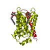
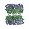
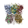
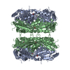
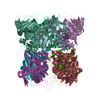
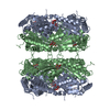
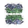
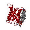
Electron crystallographic structure of lens Aquaporin-0 (AQP0) (lens MIP) in E. coli polar lipids
Components
Lens fiber major intrinsic protein
Keywords
MEMBRANE PROTEIN / water channel / lens / lipid-protein interactions
Function / homology
Function and homology information
gap junction-mediated intercellular transport / water transport / water channel activity / structural constituent of eye lens / gap junction / lens development in camera-type eye / positive regulation of cell adhesion / visual perception / protein homotetramerization / calmodulin binding ...gap junction-mediated intercellular transport / water transport / water channel activity / structural constituent of eye lens / gap junction / lens development in camera-type eye / positive regulation of cell adhesion / visual perception / protein homotetramerization / calmodulin binding / apical plasma membrane / endoplasmic reticulum / plasma membrane Similarity search - Function
Glycerol uptake facilitator protein / Glycerol uptake facilitator protein. / Aquaporin transporter / Major intrinsic protein, conserved site / MIP family signature. / Major intrinsic protein / Major intrinsic protein / Aquaporin-like / Up-down Bundle / Mainly Alpha Similarity search - Domain/homology
Journal: EMBO J / Year: 2010 Title: Principles of membrane protein interactions with annular lipids deduced from aquaporin-0 2D crystals. Authors: Richard K Hite / Zongli Li / Thomas Walz / Abstract: We have previously described the interactions of aquaporin-0 (AQP0) with dimyristoyl phosphatidylcholine (DMPC) lipids. We have now determined the 2.5 A structure of AQP0 in two-dimensional (2D) ...We have previously described the interactions of aquaporin-0 (AQP0) with dimyristoyl phosphatidylcholine (DMPC) lipids. We have now determined the 2.5 A structure of AQP0 in two-dimensional (2D) crystals formed with Escherichia coli polar lipids (EPLs), which differ from DMPC both in headgroups and acyl chains. Comparison of the two structures shows that AQP0 does not adapt to the different length of the acyl chains in EPLs and that the distance between the phosphodiester groups in the two leaflets of the DMPC and EPL bilayers is almost identical. The EPL headgroups interact differently with AQP0 than do those of DMPC, but the acyl chains in the EPL and DMPC bilayers occupy similar positions. The interactions of annular lipids with membrane proteins seem to be driven by the propensity of the acyl chains to fill gaps in the protein surface. Interactions of the lipid headgroups may be responsible for the specific interactions found in tightly bound lipids but seem to have a negligible effect on interactions of generic annular lipids with membrane proteins.
History
Deposition
Mar 22, 2010
Deposition site: RCSB / Processing site: RCSB
Revision 1.0
May 12, 2010
Provider: repository / Type: Initial release
Revision 1.1
Jul 13, 2011
Group: Version format compliance
Revision 1.2
Nov 8, 2017
Group: Data collection / Data processing / Refinement description Category: em_3d_reconstruction / em_image_scans / software
Mass: 18.015 Da / Num. of mol.: 8 / Source method: isolated from a natural source / Formula: H2O
-
Experimental details
-
Experiment
Experiment
Method: ELECTRON CRYSTALLOGRAPHY / Number of used crystals: 281
EM experiment
Aggregation state: 2D ARRAY / 3D reconstruction method: electron crystallography
-
Sample preparation
Component
Name: lens Aquaporin 0 / Type: COMPLEX
Buffer solution
pH: 8
Specimen
Embedding applied: NO / Shadowing applied: NO / Staining applied: NO / Vitrification applied: YES Details: Purified membranes were solubilized in 4% (w/v) octyl glucoside in 10 mM Tris (pH 8.0) for 30 min at 22C
Crystal grow
Temperature: 300 K / Method: microdialysis / pH: 6 Details: 100 mM NaCl, 50mM MgCl2, 10mM MES pH 6.0, MICRODIALYSIS, temperature 300K
-
Data collection
Experimental equipment
Model: Tecnai Polara / Image courtesy: FEI Company
Microscopy
Model: FEI POLARA 300
Electron gun
Electron source: FIELD EMISSION GUN / Accelerating voltage: 300 kV / Illumination mode: FLOOD BEAM
In the structure databanks used in Yorodumi, some data are registered as the other names, "COVID-19 virus" and "2019-nCoV". Here are the details of the virus and the list of structure data.
Jan 31, 2019. EMDB accession codes are about to change! (news from PDBe EMDB page)
EMDB accession codes are about to change! (news from PDBe EMDB page)
The allocation of 4 digits for EMDB accession codes will soon come to an end. Whilst these codes will remain in use, new EMDB accession codes will include an additional digit and will expand incrementally as the available range of codes is exhausted. The current 4-digit format prefixed with “EMD-” (i.e. EMD-XXXX) will advance to a 5-digit format (i.e. EMD-XXXXX), and so on. It is currently estimated that the 4-digit codes will be depleted around Spring 2019, at which point the 5-digit format will come into force.
The EM Navigator/Yorodumi systems omit the EMD- prefix.
Related info.:Q: What is EMD? / ID/Accession-code notation in Yorodumi/EM Navigator
Yorodumi is a browser for structure data from EMDB, PDB, SASBDB, etc.
This page is also the successor to EM Navigator detail page, and also detail information page/front-end page for Omokage search.
The word "yorodu" (or yorozu) is an old Japanese word meaning "ten thousand". "mi" (miru) is to see.
Related info.:EMDB / PDB / SASBDB / Comparison of 3 databanks / Yorodumi Search / Aug 31, 2016. New EM Navigator & Yorodumi / Yorodumi Papers / Jmol/JSmol / Function and homology information / Changes in new EM Navigator and Yorodumi
 Movie
Movie Controller

 Yorodumi
 Open data
 Basic information
 Components
 Keywords
 Function and homology information
 MOLECULAR REPLACEMENT / cryo EM / Resolution: 2.5 Å
  Authors
 Citation
 Structure visualization
 Downloads & links
 Other downloads


 PDBj
PDBj
 Assembly

 Mass: 748.065 Da / Num. of mol.: 7 / Source method: obtained synthetically / Formula: C41H82NO8P / Comment: phospholipid*YM
 Mass: 18.015 Da / Num. of mol.: 8 / Source method: isolated from a natural source / Formula: H2O
 Sample preparation
 Processing