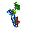
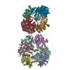
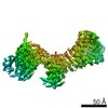
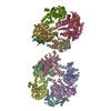
Journal: Proc Natl Acad Sci U S A / Year: 2015 Title: Electron crystallography of ultrathin 3D protein crystals: atomic model with charges. Authors: Koji Yonekura / Kazuyuki Kato / Mitsuo Ogasawara / Masahiro Tomita / Chikashi Toyoshima / Abstract: Membrane proteins and macromolecular complexes often yield crystals too small or too thin for even the modern synchrotron X-ray beam. Electron crystallography could provide a powerful means for ...Membrane proteins and macromolecular complexes often yield crystals too small or too thin for even the modern synchrotron X-ray beam. Electron crystallography could provide a powerful means for structure determination with such undersized crystals, as protein atoms diffract electrons four to five orders of magnitude more strongly than they do X-rays. Furthermore, as electron crystallography yields Coulomb potential maps rather than electron density maps, it could provide a unique method to visualize the charged states of amino acid residues and metals. Here we describe an attempt to develop a methodology for electron crystallography of ultrathin (only a few layers thick) 3D protein crystals and present the Coulomb potential maps at 3.4-Å and 3.2-Å resolution, respectively, obtained from Ca(2+)-ATPase and catalase crystals. These maps demonstrate that it is indeed possible to build atomic models from such crystals and even to determine the charged states of amino acid residues in the Ca(2+)-binding sites of Ca(2+)-ATPase and that of the iron atom in the heme in catalase.
History
Deposition
Aug 7, 2014
Deposition site: RCSB / Processing site: PDBJ
Revision 1.0
Feb 18, 2015
Provider: repository / Type: Initial release
Revision 1.1
Sep 28, 2016
Group: Database references
-
Structure visualization
Movie
Biological unit as author_and_software_defined_assembly
Mass: 18.015 Da / Num. of mol.: 155 / Source method: isolated from a natural source / Formula: H2O
Sequence details
RESIDUE 994 G IN THIS STRUCTURE IS NATURAL VARIATION ACCORDING TO DATABASE P04191-2 (AT2A1_RABIT) ...RESIDUE 994 G IN THIS STRUCTURE IS NATURAL VARIATION ACCORDING TO DATABASE P04191-2 (AT2A1_RABIT) ISOFORM SERCA1A. C-TERMINAL RESIDUES 994-1001 (DPEDERRK) ARE REPLACED BY G IN ISOFORM SERCA1A.
-
Experimental details
-
Experiment
Experiment
Method: ELECTRON CRYSTALLOGRAPHY
EM experiment
Aggregation state: 3D ARRAY / 3D reconstruction method: electron crystallography
-
Sample preparation
Component
Name: calcium ATPase / Type: COMPLEX
Specimen
Embedding applied: NO / Shadowing applied: NO / Staining applied: NO / Vitrification applied: YES
Vitrification
Cryogen name: ETHANE
-
Data collection
Microscopy
Model: HITACHI HF3000
Electron gun
Electron source: FIELD EMISSION GUN / Accelerating voltage: 300 kV / Illumination mode: FLOOD BEAM
Electron lens
Mode: DIFFRACTION
Image recording
Film or detector model: TVIPS TEMCAM-F224 (2k x 2k)
Resolution: 3.4→8 Å / Cor.coef. Fo:Fc: 0.798 / Cor.coef. Fo:Fc free: 0.768 / SU B: 27.539 / SU ML: 0.5 / Cross valid method: THROUGHOUT / σ(F): 0 / ESU R Free: 0.939 / Stereochemistry target values: MAXIMUM LIKELIHOOD Details: HYDROGENS HAVE BEEN ADDED IN THE RIDING POSITIONS U VALUES: REFINED INDIVIDUALLY
Rfactor
Num. reflection
% reflection
Selection details
Rfree
0.3152
436
3 %
RANDOM
Rwork
0.2769
14184
-
-
obs
0.278
-
67.48 %
-
Solvent computation
Ion probe radii: 0.8 Å / Shrinkage radii: 0.8 Å / VDW probe radii: 1.2 Å / Solvent model: BABINET MODEL WITH MASK
In the structure databanks used in Yorodumi, some data are registered as the other names, "COVID-19 virus" and "2019-nCoV". Here are the details of the virus and the list of structure data.
Jan 31, 2019. EMDB accession codes are about to change! (news from PDBe EMDB page)
EMDB accession codes are about to change! (news from PDBe EMDB page)
The allocation of 4 digits for EMDB accession codes will soon come to an end. Whilst these codes will remain in use, new EMDB accession codes will include an additional digit and will expand incrementally as the available range of codes is exhausted. The current 4-digit format prefixed with “EMD-” (i.e. EMD-XXXX) will advance to a 5-digit format (i.e. EMD-XXXXX), and so on. It is currently estimated that the 4-digit codes will be depleted around Spring 2019, at which point the 5-digit format will come into force.
The EM Navigator/Yorodumi systems omit the EMD- prefix.
Related info.:Q: What is EMD? / ID/Accession-code notation in Yorodumi/EM Navigator
Yorodumi is a browser for structure data from EMDB, PDB, SASBDB, etc.
This page is also the successor to EM Navigator detail page, and also detail information page/front-end page for Omokage search.
The word "yorodu" (or yorozu) is an old Japanese word meaning "ten thousand". "mi" (miru) is to see.
Related info.:EMDB / PDB / SASBDB / Comparison of 3 databanks / Yorodumi Search / Aug 31, 2016. New EM Navigator & Yorodumi / Yorodumi Papers / Jmol/JSmol / Function and homology information / Changes in new EM Navigator and Yorodumi
 Movie
Movie Controller

 Yorodumi
 Open data
 Basic information
 Components
 Keywords
 Function and homology information
 Authors
 Citation
 Structure visualization
 Downloads & links
 Other downloads
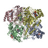


 PDBj
PDBj


 Assembly
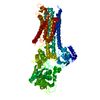


 Mass: 40.078 Da / Num. of mol.: 2 / Source method: obtained synthetically / Formula: Ca
 Mass: 22.990 Da / Num. of mol.: 1 / Source method: obtained synthetically / Formula: Na
 Mass: 18.015 Da / Num. of mol.: 155 / Source method: isolated from a natural source / Formula: H2O
 Sample preparation
 FIELD EMISSION GUN / Accelerating voltage: 300 kV / Illumination mode: FLOOD BEAM
 Processing