 Movie
Movie Controller


+ Open data

- Basic information
| Entry | Database: EMDB / ID: EMD-5945 | |||||||||
|---|---|---|---|---|---|---|---|---|---|---|
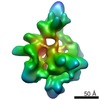
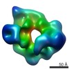
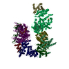
| Title | Pyruvate Carboxylase tetramer in asymmetric architecture | |||||||||
 Map data Map data | CryoEM of tetrameric Pyruvate Carboxylase in asymmetric architecture. Calculated after sorting of a catalytically active population. | |||||||||
 Sample Sample |
| |||||||||
 Keywords Keywords | enzyme / tetrameric / biotin carboxylase | |||||||||
| Biological species |   Staphylococcus aureus (bacteria) Staphylococcus aureus (bacteria) | |||||||||
| Method | single particle reconstruction / cryo EM / Resolution: 11.5 Å | |||||||||
 Authors Authors | Lasso G / Yu LPC / Gil D / Lazaro M / Tong L / Valle M | |||||||||
 Citation Citation | Journal: Structure / Year: 2014 Title: Functional conformations for pyruvate carboxylase during catalysis explored by cryoelectron microscopy. Authors: Gorka Lasso / Linda P C Yu / David Gil / Melisa Lázaro / Liang Tong / Mikel Valle /   Abstract: The tetrameric enzyme pyruvate carboxylase (PC), a biotin-dependent carboxylase, produces oxaloacetate by two consecutive reactions that take place in distant active sites. Previous crystal ...The tetrameric enzyme pyruvate carboxylase (PC), a biotin-dependent carboxylase, produces oxaloacetate by two consecutive reactions that take place in distant active sites. Previous crystal structures revealed two different configurations for PC tetramers, the so-called symmetric and asymmetric, which were understood as characteristic molecular architectures for PC from different organisms. We have analyzed PC samples from Staphylococcus aureus while the enzyme generates oxaloacetate, expecting PC tetramers to display the conformational landscape relevant for its functioning. Using cryoelectron microscopy (cryo-EM) and sorting techniques, we detect previously defined symmetric and asymmetric architectures, demonstrating that PC maps both arrangements by large conformational changes. Furthermore, we observe that each configuration is coupled to one of the two consecutive enzymatic reactions. The findings describe the structural transitions relevant for the allosteric control of the multifunctional PC and demonstrate that by cryo-EM and classification, we can characterize freely working macromolecules. | |||||||||
| History |
|
- Structure visualization
| Movie |
 Movie viewer Movie viewer |
|---|---|
| Structure viewer | EM map: SurfViewMolmilJmol/JSmol |
| Supplemental images |

- Downloads & links
-EMDB archive
| Map data | emd_5945.map.gz | 2.5 MB | EMDB map data format | |
|---|---|---|---|---|
| Header (meta data) | emd-5945-v30.xmlemd-5945.xml | 9.9 KB 9.9 KB | Display Display | EMDB header |
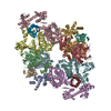
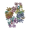
| Images | 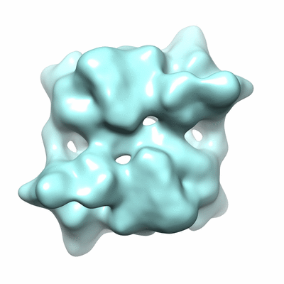 400_5945.gif 400_5945.gif 80_5945.gif 80_5945.gif | 49.6 KB 3.9 KB | ||
| Archive directory |  http://ftp.pdbj.org/pub/emdb/structures/EMD-5945ftp://ftp.pdbj.org/pub/emdb/structures/EMD-5945 http://ftp.pdbj.org/pub/emdb/structures/EMD-5945ftp://ftp.pdbj.org/pub/emdb/structures/EMD-5945 | HTTPS FTP |
-Validation report
| Summary document | emd_5945_validation.pdf.gz | 78.6 KB | Display | EMDB validaton report |
|---|---|---|---|---|
| Full document | emd_5945_full_validation.pdf.gz | 77.7 KB | Display | |
| Data in XML | emd_5945_validation.xml.gz | 494 B | Display | |
| Arichive directory | https://ftp.pdbj.org/pub/emdb/validation_reports/EMD-5945ftp://ftp.pdbj.org/pub/emdb/validation_reports/EMD-5945 | HTTPS FTP |
-Related structure data


-Links
| EMDB pages | EMDB (EBI/PDBe) / EMDataResource |
|---|
-Map
| File | Download / File: emd_5945.map.gz / Format: CCP4 / Size: 25.6 MB / Type: IMAGE STORED AS FLOATING POINT NUMBER (4 BYTES) | ||||||||||||||||||||||||||||||||||||||||||||||||||||||||||||||||||||
|---|---|---|---|---|---|---|---|---|---|---|---|---|---|---|---|---|---|---|---|---|---|---|---|---|---|---|---|---|---|---|---|---|---|---|---|---|---|---|---|---|---|---|---|---|---|---|---|---|---|---|---|---|---|---|---|---|---|---|---|---|---|---|---|---|---|---|---|---|---|
| Annotation | CryoEM of tetrameric Pyruvate Carboxylase in asymmetric architecture. Calculated after sorting of a catalytically active population. | ||||||||||||||||||||||||||||||||||||||||||||||||||||||||||||||||||||
| Voxel size | X=Y=Z: 1.75 Å | ||||||||||||||||||||||||||||||||||||||||||||||||||||||||||||||||||||
| Density |
| ||||||||||||||||||||||||||||||||||||||||||||||||||||||||||||||||||||
| Symmetry | Space group: 1 | ||||||||||||||||||||||||||||||||||||||||||||||||||||||||||||||||||||
| Details | EMDB XML:
CCP4 map header:
| ||||||||||||||||||||||||||||||||||||||||||||||||||||||||||||||||||||
-Supplemental data
- Sample components
-Entire : Pyruvate Carboxylase
| Entire | Name: Pyruvate Carboxylase |
|---|---|
| Components |
|
-Supramolecule #1000: Pyruvate Carboxylase
| Supramolecule | Name: Pyruvate Carboxylase / type: sample / ID: 1000 / Oligomeric state: Homotetramer / Number unique components: 1 |
|---|---|
| Molecular weight | Theoretical: 520 KDa |
-Macromolecule #1: pyruvate carboxylate
| Macromolecule | Name: pyruvate carboxylate / type: protein_or_peptide / ID: 1 / Number of copies: 4 / Oligomeric state: tetramer / Recombinant expression: Yes |
|---|---|
| Source (natural) | Organism: Staphylococcus aureus (bacteria) |
| Molecular weight | Theoretical: 520 KDa |
| Recombinant expression | Organism: |
-Experimental details
-Structure determination
| Method | cryo EM |
|---|---|
 Processing Processing | single particle reconstruction |
| Aggregation state | particle |
-Sample preparation
| Concentration | 0.05 mg/mL |
|---|---|
| Buffer | pH: 7.5 Details: 16.6 mM Tris-HCl, 1.7 mM acetyl-CoA, 15.7 mM pyruvate, 4.1 mM MgCl2, 1.7 mM DTT, 166.6 mM NaCl, 8.3 mM KHCO3, 1.66 mM ATP. |
| Vitrification | Cryogen name: ETHANE / Chamber humidity: 95 % / Instrument: FEI VITROBOT MARK II |
- Electron microscopy
| Microscope | JEOL 2200FSC |
|---|---|
| Specialist optics | Energy filter - Name: Omega in-column |
| Date | Jan 1, 2011 |
| Image recording | Category: FILM / Film or detector model: KODAK SO-163 FILM / Digitization - Scanner: ZEISS SCAI / Digitization - Sampling interval: 7 µm / Number real images: 50 / Average electron dose: 15 e/Å2 / Bits/pixel: 16 |
| Electron beam | Acceleration voltage: 200 kV / Electron source:  FIELD EMISSION GUN FIELD EMISSION GUN |
| Electron optics | Illumination mode: FLOOD BEAM / Imaging mode: BRIGHT FIELD / Cs: 2.0 mm / Nominal magnification: 40000 |
| Sample stage | Specimen holder model: GATAN LIQUID NITROGEN |
-Image processing
| CTF correction | Details: Volume correction in defocus groups |
|---|---|
| Final reconstruction | Algorithm: OTHER / Resolution.type: BY AUTHOR / Resolution: 11.5 Å / Resolution method: OTHER / Software - Name: Relion, Spider / Number images used: 14032 |
-Atomic model buiding 1
| Initial model | PDB ID: |
|---|---|
| Software | Name: MDFF |
| Refinement | Space: REAL / Protocol: FLEXIBLE FIT |

