 Movie
Movie Controller

[English] 日本語
 Yorodumi
- EMDB-5466: Cryo-electron microscopy reconstruction of the Enterovirus 71 emp... -
+ Open data

- Basic information
| Entry | Database: EMDB / ID: EMD-5466 | |||||||||
|---|---|---|---|---|---|---|---|---|---|---|
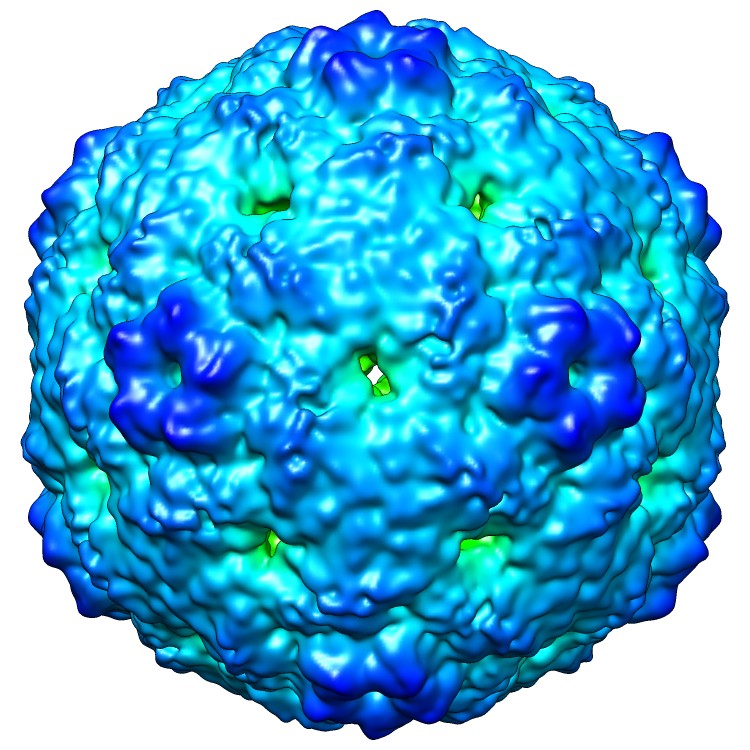
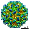
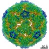
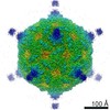
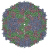
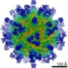
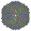
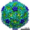
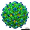
| Title | Cryo-electron microscopy reconstruction of the Enterovirus 71 empty capsid | |||||||||
 Map data Map data | Reconstruction of the EV71 empty capsid. | |||||||||
 Sample Sample |
| |||||||||
 Keywords Keywords | Enterovirus 71 / EV71 / empty capsid / 80S | |||||||||
| Function / homology |  Function and homology information Function and homology informationsymbiont-mediated suppression of host cytoplasmic pattern recognition receptor signaling pathway via inhibition of MDA-5 activity / picornain 2A / symbiont-mediated suppression of host mRNA export from nucleus / symbiont genome entry into host cell via pore formation in plasma membrane / picornain 3C / T=pseudo3 icosahedral viral capsid / host cell cytoplasmic vesicle membrane / cytoplasmic vesicle membrane / endocytosis involved in viral entry into host cell / nucleoside-triphosphate phosphatase ...symbiont-mediated suppression of host cytoplasmic pattern recognition receptor signaling pathway via inhibition of MDA-5 activity / picornain 2A / symbiont-mediated suppression of host mRNA export from nucleus / symbiont genome entry into host cell via pore formation in plasma membrane / picornain 3C / T=pseudo3 icosahedral viral capsid / host cell cytoplasmic vesicle membrane / cytoplasmic vesicle membrane / endocytosis involved in viral entry into host cell / nucleoside-triphosphate phosphatase / protein complex oligomerization / monoatomic ion channel activity / RNA helicase activity / induction by virus of host autophagy / cysteine-type endopeptidase activity / RNA-directed RNA polymerase / viral RNA genome replication / virus-mediated perturbation of host defense response / RNA-dependent RNA polymerase activity / DNA-templated transcription / host cell nucleus / virion attachment to host cell / structural molecule activity / ATP hydrolysis activity / proteolysis / RNA binding / ATP binding / metal ion binding Similarity search - Function | |||||||||
| Biological species |   Human enterovirus 71 Human enterovirus 71 | |||||||||
| Method | single particle reconstruction / cryo EM / Resolution: 9.2 Å | |||||||||
 Authors Authors | Shingler KL / Yoder JL / Carnegie MS / Ashley RE / Makhov AM / Conway JF / Hafenstein S | |||||||||
 Citation Citation | Journal: PLoS Pathog / Year: 2013 Title: The enterovirus 71 A-particle forms a gateway to allow genome release: a cryoEM study of picornavirus uncoating. Authors: Kristin L Shingler / Jennifer L Yoder / Michael S Carnegie / Robert E Ashley / Alexander M Makhov / James F Conway / Susan Hafenstein /  Abstract: Since its discovery in 1969, enterovirus 71 (EV71) has emerged as a serious worldwide health threat. This human pathogen of the picornavirus family causes hand, foot, and mouth disease, and also has ...Since its discovery in 1969, enterovirus 71 (EV71) has emerged as a serious worldwide health threat. This human pathogen of the picornavirus family causes hand, foot, and mouth disease, and also has the capacity to invade the central nervous system to cause severe disease and death. Upon binding to a host receptor on the cell surface, the virus begins a two-step uncoating process, first forming an expanded, altered "A-particle", which is primed for genome release. In a second step after endocytosis, an unknown trigger leads to RNA expulsion, generating an intact, empty capsid. Cryo-electron microscopy reconstructions of these two capsid states provide insight into the mechanics of genome release. The EV71 A-particle capsid interacts with the genome near the icosahedral two-fold axis of symmetry, which opens to the external environment via a channel ∼10 Å in diameter that is lined with patches of negatively charged residues. After the EV71 genome has been released, the two-fold channel shrinks, though the overall capsid dimensions are conserved. These structural characteristics identify the two-fold channel as the site where a gateway forms and regulates the process of genome release. | |||||||||
| History |
|
- Structure visualization
| Movie |
Movie viewer |
|---|---|
| Structure viewer | EM map: SurfViewMolmilJmol/JSmol |
| Supplemental images |
- Downloads & links
-EMDB archive
| Map data | emd_5466.map.gz | 62.3 MB | EMDB map data format | |
|---|---|---|---|---|
| Header (meta data) | emd-5466-v30.xmlemd-5466.xml | 11.7 KB 11.7 KB | Display Display | EMDB header |
| Images |  emd_5466_1.jpg emd_5466_1.jpg | 142 KB | ||
| Archive directory |  http://ftp.pdbj.org/pub/emdb/structures/EMD-5466ftp://ftp.pdbj.org/pub/emdb/structures/EMD-5466 http://ftp.pdbj.org/pub/emdb/structures/EMD-5466ftp://ftp.pdbj.org/pub/emdb/structures/EMD-5466 | HTTPS FTP |
-Validation report
| Summary document | emd_5466_validation.pdf.gz | 360 KB | Display | EMDB validaton report |
|---|---|---|---|---|
| Full document | emd_5466_full_validation.pdf.gz | 359.5 KB | Display | |
| Data in XML | emd_5466_validation.xml.gz | 7 KB | Display | |
| Arichive directory | https://ftp.pdbj.org/pub/emdb/validation_reports/EMD-5466ftp://ftp.pdbj.org/pub/emdb/validation_reports/EMD-5466 | HTTPS FTP |
-Related structure data
| Related structure data |  3j23MC  5465C  3j22C M: atomic model generated by this map C: citing same article ( |
|---|---|
| Similar structure data |
-Links
| EMDB pages | EMDB (EBI/PDBe) / EMDataResource |
|---|---|
| Related items in Molecule of the Month |


-Map
| File | Download / File: emd_5466.map.gz / Format: CCP4 / Size: 147.7 MB / Type: IMAGE STORED AS FLOATING POINT NUMBER (4 BYTES) | ||||||||||||||||||||||||||||||||||||||||||||||||||||||||||||||||||||
|---|---|---|---|---|---|---|---|---|---|---|---|---|---|---|---|---|---|---|---|---|---|---|---|---|---|---|---|---|---|---|---|---|---|---|---|---|---|---|---|---|---|---|---|---|---|---|---|---|---|---|---|---|---|---|---|---|---|---|---|---|---|---|---|---|---|---|---|---|---|
| Annotation | Reconstruction of the EV71 empty capsid. | ||||||||||||||||||||||||||||||||||||||||||||||||||||||||||||||||||||
| Voxel size | X=Y=Z: 1.27 Å | ||||||||||||||||||||||||||||||||||||||||||||||||||||||||||||||||||||
| Density |
| ||||||||||||||||||||||||||||||||||||||||||||||||||||||||||||||||||||
| Symmetry | Space group: 1 | ||||||||||||||||||||||||||||||||||||||||||||||||||||||||||||||||||||
| Details | EMDB XML:
CCP4 map header:
| ||||||||||||||||||||||||||||||||||||||||||||||||||||||||||||||||||||
-Supplemental data
- Sample components
-Entire : Enterovirus 71 empty capsid
| Entire | Name: Enterovirus 71 empty capsid |
|---|---|
| Components |
|
-Supramolecule #1000: Enterovirus 71 empty capsid
| Supramolecule | Name: Enterovirus 71 empty capsid / type: sample / ID: 1000 Details: The sample was purified mature virus heated in solution. Oligomeric state: icosahedral virus / Number unique components: 1 |
|---|---|
| Molecular weight | Experimental: 7 MDa / Theoretical: 7 MDa / Method: calculation |
-Supramolecule #1: Human enterovirus 71
| Supramolecule | Name: Human enterovirus 71 / type: virus / ID: 1 / Name.synonym: EV71, Hand foot and mouth disease virus / Details: purified virus in solution / NCBI-ID: 39054 / Sci species name: Human enterovirus 71 / Database: NCBI / Virus type: VIRION / Virus isolate: SEROTYPE / Virus enveloped: No / Virus empty: Yes / Syn species name: EV71, Hand foot and mouth disease virus |
|---|---|
| Host (natural) | Organism:  Homo sapiens (human) / synonym: VERTEBRATES Homo sapiens (human) / synonym: VERTEBRATES |
| Molecular weight | Experimental: 7 MDa / Theoretical: 7 MDa |
| Virus shell | Shell ID: 1 / Name: single capsid shell / Diameter: 300 Å / T number (triangulation number): 1 |
-Experimental details
-Structure determination
| Method | cryo EM |
|---|---|
 Processing Processing | single particle reconstruction |
| Aggregation state | particle |
-Sample preparation
| Concentration | 0.1 mg/mL |
|---|---|
| Buffer | pH: 7.5 / Details: 10 mM Tris-HCl, 20 mM NaCl, 5 mM MgCl2 |
| Grid | Details: glow discharged holey carbon Quantifoil electron microscopy grids |
| Vitrification | Cryogen name: ETHANE-PROPANE MIXTURE / Chamber humidity: 95 % / Chamber temperature: 77 K / Instrument: FEI VITROBOT MARK III |
- Electron microscopy
| Microscope | FEI TECNAI F20 |
|---|---|
| Temperature | Average: 95 K |
| Alignment procedure | Legacy - Astigmatism: CTFFIND3 (EMAN) |
| Date | Apr 17, 2012 |
| Image recording | Category: FILM / Film or detector model: KODAK SO-163 FILM / Digitization - Scanner: NIKON SUPER COOLSCAN 9000 / Digitization - Sampling interval: 6.35 µm / Number real images: 40 / Average electron dose: 15 e/Å2 |
| Electron beam | Acceleration voltage: 200 kV / Electron source:  FIELD EMISSION GUN FIELD EMISSION GUN |
| Electron optics | Calibrated magnification: 50000 / Illumination mode: FLOOD BEAM / Imaging mode: BRIGHT FIELD / Cs: 2 mm / Nominal defocus max: 4.37 µm / Nominal defocus min: 1.73 µm / Nominal magnification: 50000 |
| Sample stage | Specimen holder model: GATAN LIQUID NITROGEN |
| Experimental equipment |  Model: Tecnai F20 / Image courtesy: FEI Company |
-Image processing
| Details | These particles were selected individually. |
|---|---|
| CTF correction | Details: auto3DEM |
| Final reconstruction | Algorithm: OTHER / Resolution.type: BY AUTHOR / Resolution: 9.2 Å / Resolution method: FSC 0.5 CUT-OFF / Software - Name: EMAN2, auto3DEM / Details: A single data set was used for the reconstruction. / Number images used: 1931 |
-Atomic model buiding 1
| Initial model | PDB ID: Chain - #0 - Chain ID: A / Chain - #1 - Chain ID: B / Chain - #2 - Chain ID: C |
|---|---|
| Software | Name: Chimera, Situs |
| Details | Protocol: rigid body. The protomer was fit in Chimera, used to generate a full crystal map, then docked as a rigid body in Situs. |
| Refinement | Space: REAL / Protocol: RIGID BODY FIT / Target criteria: correlation coefficient |
| Output model | PDB-3j23: |

