 Movie
Movie Controller

[English] 日本語
 Yorodumi
- EMDB-1870: Three-Dimensional Reconstruction of Heterocapsa circularisquama R... -
+ Open data

- Basic information
| Entry | Database: EMDB / ID: EMD-1870 | |||||||||
|---|---|---|---|---|---|---|---|---|---|---|
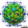
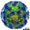
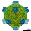
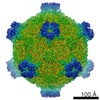
| Title | Three-Dimensional Reconstruction of Heterocapsa circularisquama RNA Virus (HcRNAV-109) by Cryo-Electron Microscopy | |||||||||
 Map data Map data | Heterocapsa circularisquama ss RNA Virus (HcRNAV-109) | |||||||||
 Sample Sample |
| |||||||||
 Keywords Keywords | Marine Virus / single-stranded RNA Virus / handedness determination / Alvernaviridae / Dinornavirus | |||||||||
| Biological species |  Heterocapsa circularisquama (eukaryote) / Heterocapsa circularisquama (eukaryote) /  Heterocapsa circularisquama RNA virus Heterocapsa circularisquama RNA virus | |||||||||
| Method | single particle reconstruction / cryo EM / negative staining / Resolution: 18.0 Å | |||||||||
 Authors Authors | Miller JL / Woodward JD / Chen S / Jaffer MA / Weber BW / Nagasaki K / Tomaru Y / Wepf R / Roseman A / Sewell BT / Varsani A | |||||||||
 Citation Citation | Journal: J Gen Virol / Year: 2011 Title: Three-dimensional reconstruction of Heterocapsa circularisquama RNA virus by electron cryo-microscopy. Authors: Jennifer L Miller / Jeremy Woodward / Shaoxia Chen / Mohammed Jaffer / Brandon Weber / Keizo Nagasaki / Yuji Tomaru / Roger Wepf / Alan Roseman / Arvind Varsani / Trevor Sewell /      Abstract: Heterocapsa circularisquama RNA virus is a non-enveloped icosahedral ssRNA virus infectious to the harmful bloom-forming dinoflagellate, H. circularisquama, and which is assumed to be the major ...Heterocapsa circularisquama RNA virus is a non-enveloped icosahedral ssRNA virus infectious to the harmful bloom-forming dinoflagellate, H. circularisquama, and which is assumed to be the major natural agent controlling the host population. The viral capsid is constructed from a single gene product. Electron cryo-microscopy revealed that the virus has a diameter of 34 nm and T = 3 symmetry. The 180 quasi-equivalent monomers have an unusual arrangement in that each monomer contributes to a 'bump' on the surface of the protein. Though the capsid protein probably has the classic 'jelly roll' β-sandwich fold, this is a new packing arrangement and is distantly related to the other positive-sense ssRNA virus capsid proteins. The handedness of the structure has been determined by a novel method involving high resolution scanning electron microscopy of the negatively stained viruses and secondary electron detection. | |||||||||
| History |
|
- Structure visualization
| Movie |
 Movie viewer Movie viewer |
|---|---|
| Structure viewer | EM map: SurfViewMolmilJmol/JSmol |
| Supplemental images |

- Downloads & links
-EMDB archive
| Map data | emd_1870.map.gz | 28.7 MB | EMDB map data format | |
|---|---|---|---|---|
| Header (meta data) | emd-1870-v30.xmlemd-1870.xml | 10.6 KB 10.6 KB | Display Display | EMDB header |
| Images | 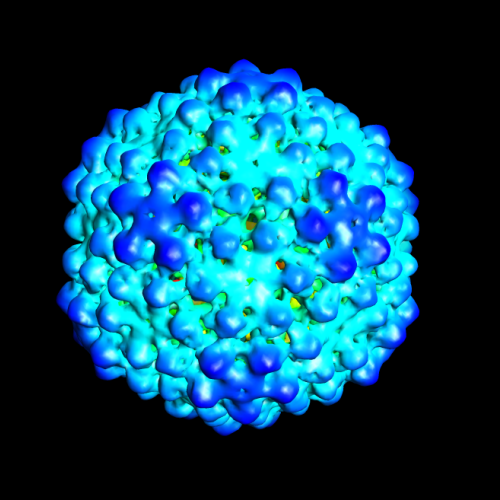 1870.png 1870.png | 234.3 KB | ||
| Archive directory |  http://ftp.pdbj.org/pub/emdb/structures/EMD-1870ftp://ftp.pdbj.org/pub/emdb/structures/EMD-1870 http://ftp.pdbj.org/pub/emdb/structures/EMD-1870ftp://ftp.pdbj.org/pub/emdb/structures/EMD-1870 | HTTPS FTP |
-Validation report
| Summary document | emd_1870_validation.pdf.gz | 275.1 KB | Display | EMDB validaton report |
|---|---|---|---|---|
| Full document | emd_1870_full_validation.pdf.gz | 274.2 KB | Display | |
| Data in XML | emd_1870_validation.xml.gz | 6 KB | Display | |
| Arichive directory | https://ftp.pdbj.org/pub/emdb/validation_reports/EMD-1870ftp://ftp.pdbj.org/pub/emdb/validation_reports/EMD-1870 | HTTPS FTP |
-Related structure data
| Similar structure data |
|---|

-Links
| EMDB pages | EMDB (EBI/PDBe) / EMDataResource |
|---|
-Map
| File | Download / File: emd_1870.map.gz / Format: CCP4 / Size: 29.8 MB / Type: IMAGE STORED AS FLOATING POINT NUMBER (4 BYTES) | ||||||||||||||||||||||||||||||||||||||||||||||||||||||||||||||||||||
|---|---|---|---|---|---|---|---|---|---|---|---|---|---|---|---|---|---|---|---|---|---|---|---|---|---|---|---|---|---|---|---|---|---|---|---|---|---|---|---|---|---|---|---|---|---|---|---|---|---|---|---|---|---|---|---|---|---|---|---|---|---|---|---|---|---|---|---|---|---|
| Annotation | Heterocapsa circularisquama ss RNA Virus (HcRNAV-109) | ||||||||||||||||||||||||||||||||||||||||||||||||||||||||||||||||||||
| Voxel size | X=Y=Z: 3.616 Å | ||||||||||||||||||||||||||||||||||||||||||||||||||||||||||||||||||||
| Density |
| ||||||||||||||||||||||||||||||||||||||||||||||||||||||||||||||||||||
| Symmetry | Space group: 1 | ||||||||||||||||||||||||||||||||||||||||||||||||||||||||||||||||||||
| Details | EMDB XML:
CCP4 map header:
| ||||||||||||||||||||||||||||||||||||||||||||||||||||||||||||||||||||
-Supplemental data
- Sample components
-Entire : Heterocapsa circularisquama RNA virus (HcRNAV-109)
| Entire | Name: Heterocapsa circularisquama RNA virus (HcRNAV-109) |
|---|---|
| Components |
|
-Supramolecule #1000: Heterocapsa circularisquama RNA virus (HcRNAV-109)
| Supramolecule | Name: Heterocapsa circularisquama RNA virus (HcRNAV-109) / type: sample / ID: 1000 / Details: T equals 3 Oligomeric state: Virus particle containing 180 identical protein subunits and one single-stranded RNA molecule Number unique components: 2 |
|---|
-Supramolecule #1: Heterocapsa circularisquama RNA virus
| Supramolecule | Name: Heterocapsa circularisquama RNA virus / type: virus / ID: 1 / Name.synonym: HcRNAV-109 / NCBI-ID: 331975 / Sci species name: Heterocapsa circularisquama RNA virus / Virus type: VIRION / Virus isolate: STRAIN / Virus enveloped: No / Virus empty: No / Syn species name: HcRNAV-109 |
|---|---|
| Host (natural) | Organism: Heterocapsa circularisquama (eukaryote) / synonym: ALGAE |
| Virus shell | Shell ID: 1 / Diameter: 340 Å / T number (triangulation number): 3 |
-Macromolecule #1: Singe Stranded RNA
| Macromolecule | Name: Singe Stranded RNA / type: rna / ID: 1 / Name.synonym: Singe Stranded RNA / Classification: OTHER / Structure: SINGLE STRANDED / Synthetic?: No |
|---|---|
| Source (natural) | Organism: Heterocapsa circularisquama (eukaryote) |
-Experimental details
-Structure determination
| Method | negative staining, cryo EM |
|---|---|
 Processing Processing | single particle reconstruction |
| Aggregation state | particle |
-Sample preparation
| Buffer | pH: 7.4 / Details: 10 mM Na2HPO4 and 10 mM KH2PO4 in distilled water |
|---|---|
| Staining | Type: NEGATIVE / Details: Cryo in ice |
| Grid | Details: 300 mesh copper R2/2 Quantifoil grids |
| Vitrification | Cryogen name: ETHANE / Chamber humidity: 90 % / Chamber temperature: 100 K / Instrument: HOMEMADE PLUNGER / Details: Vitrification instrument: Home-made plunger Method: Blot 3 seconds, re-apply virus, blot 2 seconds. plunge |
- Electron microscopy
| Microscope | FEI TECNAI F30 |
|---|---|
| Alignment procedure | Legacy - Astigmatism: Objective lens astigmatism was corrected at 100,000 times magnification |
| Image recording | Category: FILM / Film or detector model: KODAK SO-163 FILM / Digitization - Scanner: OTHER / Digitization - Sampling interval: 1.808 µm / Number real images: 108 / Average electron dose: 10 e/Å2 / Bits/pixel: 16 |
| Electron beam | Acceleration voltage: 300 kV / Electron source:  FIELD EMISSION GUN FIELD EMISSION GUN |
| Electron optics | Calibrated magnification: 55299.5 / Illumination mode: FLOOD BEAM / Imaging mode: BRIGHT FIELD / Cs: 2 mm / Nominal defocus max: 4.8 µm / Nominal defocus min: 1.6 µm / Nominal magnification: 59000 |
| Sample stage | Specimen holder: Side entry cryo holder Gatan 626 / Specimen holder model: GATAN LIQUID NITROGEN |
| Experimental equipment |  Model: Tecnai F30 / Image courtesy: FEI Company |
-Image processing
| Details | The concentration of virus in the sample was very low |
|---|---|
| CTF correction | Details: Each micrograph |
| Final reconstruction | Applied symmetry - Point group: I (icosahedral) / Algorithm: OTHER / Resolution.type: BY AUTHOR / Resolution: 18.0 Å / Resolution method: FSC 0.5 CUT-OFF / Software - Name: MRC, EMAN, SPIDER Details: We used two different starting models for refinement - one generated by EMAN starticos and one generated by applying the orientations determined by MRC refine Number images used: 2593 |
| Final angle assignment | Details: SPIDER |
