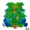
- EMDB-7403: Single-particle reconstruction of DARP14 - A designed protein sca... -
+
Open data
ID or keywords:
Loading...
-
Basic information
Entry
Database: EMDB / ID: EMD-7403
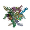
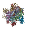
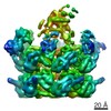
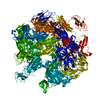
Title
Single-particle reconstruction of DARP14 - A designed protein scaffold displaying ~17kDa DARPin proteins - Helical extension
Map data
Sample
Complex: DARP14
Complex: DARP14 - Subunit A
Complex: DARP14 - Subunit B
Complex: DARP14 - DARPin
Function / homology
Function and homology information
5-carboxymethyl-2-hydroxymuconate delta-isomerase activity / corrinoid adenosyltransferase activity / : / ATP binding / metal ion binding Similarity search - Function
Journal: Proc Natl Acad Sci U S A / Year: 2018 Title: Near-atomic cryo-EM imaging of a small protein displayed on a designed scaffolding system. Authors: Yuxi Liu / Shane Gonen / Tamir Gonen / Todd O Yeates / Abstract: Current single-particle cryo-electron microscopy (cryo-EM) techniques can produce images of large protein assemblies and macromolecular complexes at atomic level detail without the need for crystal ...Current single-particle cryo-electron microscopy (cryo-EM) techniques can produce images of large protein assemblies and macromolecular complexes at atomic level detail without the need for crystal growth. However, proteins of smaller size, typical of those found throughout the cell, are not presently amenable to detailed structural elucidation by cryo-EM. Here we use protein design to create a modular, symmetrical scaffolding system to make protein molecules of typical size suitable for cryo-EM. Using a rigid continuous alpha helical linker, we connect a small 17-kDa protein (DARPin) to a protein subunit that was designed to self-assemble into a cage with cubic symmetry. We show that the resulting construct is amenable to structural analysis by single-particle cryo-EM, allowing us to identify and solve the structure of the attached small protein at near-atomic detail, ranging from 3.5- to 5-Å resolution. The result demonstrates that proteins considerably smaller than the theoretical limit of 50 kDa for cryo-EM can be visualized clearly when arrayed in a rigid fashion on a symmetric designed protein scaffold. Furthermore, because the amino acid sequence of a DARPin can be chosen to confer tight binding to various other protein or nucleic acid molecules, the system provides a future route for imaging diverse macromolecules, potentially broadening the application of cryo-EM to proteins of typical size in the cell.
History
Deposition
Jan 24, 2018
-
Header (metadata) release
Feb 28, 2018
-
Map release
Mar 21, 2018
-
Update
Apr 11, 2018
-
Current status
Apr 11, 2018
Processing site: RCSB / Status: Released
-
Structure visualization
Movie
Surface view with section colored by density value
In the structure databanks used in Yorodumi, some data are registered as the other names, "COVID-19 virus" and "2019-nCoV". Here are the details of the virus and the list of structure data.
Jan 31, 2019. EMDB accession codes are about to change! (news from PDBe EMDB page)
EMDB accession codes are about to change! (news from PDBe EMDB page)
The allocation of 4 digits for EMDB accession codes will soon come to an end. Whilst these codes will remain in use, new EMDB accession codes will include an additional digit and will expand incrementally as the available range of codes is exhausted. The current 4-digit format prefixed with “EMD-” (i.e. EMD-XXXX) will advance to a 5-digit format (i.e. EMD-XXXXX), and so on. It is currently estimated that the 4-digit codes will be depleted around Spring 2019, at which point the 5-digit format will come into force.
The EM Navigator/Yorodumi systems omit the EMD- prefix.
Related info.:Q: What is EMD? / ID/Accession-code notation in Yorodumi/EM Navigator
Yorodumi is a browser for structure data from EMDB, PDB, SASBDB, etc.
This page is also the successor to EM Navigator detail page, and also detail information page/front-end page for Omokage search.
The word "yorodu" (or yorozu) is an old Japanese word meaning "ten thousand". "mi" (miru) is to see.
Related info.:EMDB / PDB / SASBDB / Comparison of 3 databanks / Yorodumi Search / Aug 31, 2016. New EM Navigator & Yorodumi / Yorodumi Papers / Jmol/JSmol / Function and homology information / Changes in new EM Navigator and Yorodumi
 Movie
Movie Controller

 Yorodumi
 Open data
 Basic information
 Map data
 Sample
 Function and homology information
 Pyrococcus horikoshii OT3 (archaea) /
  Pseudomonas aeruginosa PAO1 (bacteria)
 Authors
 Citation
 Structure visualization
 Downloads & links
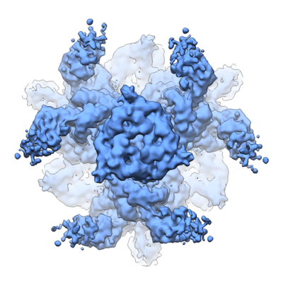 emd_7403.png
 http://ftp.pdbj.org/pub/emdb/structures/EMD-7403

 Sample components
 Processing
 Electron microscopy
 FIELD EMISSION GUN
