Movie
Movie Controller

[English] 日本語
 Yorodumi
- PDB-3deg: Complex of elongating Escherichia coli 70S ribosome and EF4(LepA)... -
+ Open data

- Basic information
| Entry | Database: PDB / ID: 3deg | ||||||
|---|---|---|---|---|---|---|---|
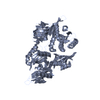
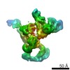
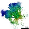
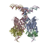
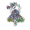
| Title | Complex of elongating Escherichia coli 70S ribosome and EF4(LepA)-GMPPNP | ||||||
 Components Components |
| ||||||
 Keywords Keywords | RIBOSOME / translation / LepA / EF4 / GTP-binding / Membrane / Nucleotide-binding / Antibiotic resistance / Ribonucleoprotein / Ribosomal protein / RNA-binding / rRNA-binding / tRNA-binding / Methylation | ||||||
| Function / homology |  Function and homology information Function and homology information: / response to pH / guanosine tetraphosphate binding / stringent response / ribosomal large subunit binding / misfolded RNA binding / Group I intron splicing / RNA folding / ribosomal small subunit binding / translation elongation factor activity ...: / response to pH / guanosine tetraphosphate binding / stringent response / ribosomal large subunit binding / misfolded RNA binding / Group I intron splicing / RNA folding / ribosomal small subunit binding / translation elongation factor activity / translational termination / response to salt stress / response to cold / positive regulation of RNA splicing / ribosomal large subunit assembly / positive regulation of translation / maintenance of translational fidelity / ribosome binding / ribosomal small subunit biogenesis / large ribosomal subunit rRNA binding / cytosolic small ribosomal subunit / cytosolic large ribosomal subunit / cytoplasmic translation / tRNA binding / rRNA binding / ribosome / structural constituent of ribosome / translation / response to antibiotic / GTPase activity / GTP binding / identical protein binding / plasma membrane / cytoplasm / cytosol Similarity search - Function | ||||||
| Biological species |  | ||||||
| Method | ELECTRON MICROSCOPY / single particle reconstruction / cryo EM / Resolution: 10.9 Å | ||||||
 Authors Authors | Connell, S.R. / Topf, M. / Qin, Y. / Wilson, D.N. / Mielke, T. / Fucini, P. / Nierhaus, K.H. / Spahn, C.M.T. | ||||||
 Citation Citation | Journal: Nat Struct Mol Biol / Year: 2008 Title: A new tRNA intermediate revealed on the ribosome during EF4-mediated back-translocation. Authors: Sean R Connell / Maya Topf / Yan Qin / Daniel N Wilson / Thorsten Mielke / Paola Fucini / Knud H Nierhaus / Christian M T Spahn /  Abstract: EF4 (LepA) is an almost universally conserved translational GTPase in eubacteria. It seems to be essential under environmental stress conditions and has previously been shown to back-translocate the ...EF4 (LepA) is an almost universally conserved translational GTPase in eubacteria. It seems to be essential under environmental stress conditions and has previously been shown to back-translocate the tRNAs on the ribosome, thereby reverting the canonical translocation reaction. In the current work, EF4 was directly visualized in the process of back-translocating tRNAs by single-particle cryo-EM. Using flexible fitting methods, we built a model of ribosome-bound EF4 based on the cryo-EM map and a recently published unbound EF4 X-ray structure. The cryo-EM map establishes EF4 as a noncanonical elongation factor that interacts not only with the elongating ribosome, but also with the back-translocated tRNA in the A-site region, which is present in a previously unseen, intermediate state and deviates markedly from the position of a canonical A-tRNA. Our results, therefore, provide insight into the underlying structural principles governing back-translocation. | ||||||
| History |
|
- Structure visualization
| Movie |
Movie viewer |
|---|---|
| Structure viewer | Molecule: MolmilJmol/JSmol |
- Downloads & links
-Download
| PDBx/mmCIF format | 3deg.cif.gz | 325.1 KB | Display | PDBx/mmCIF format |
|---|---|---|---|---|
| PDB format | pdb3deg.ent.gz | 243 KB | Display | PDB format |
| PDBx/mmJSON format | 3deg.json.gz | Tree view | PDBx/mmJSON format | |
| Others |  Other downloads Other downloads |
-Validation report
| Summary document | 3deg_validation.pdf.gz | 926.5 KB | Display | wwPDB validaton report |
|---|---|---|---|---|
| Full document | 3deg_full_validation.pdf.gz | 1020.9 KB | Display | |
| Data in XML | 3deg_validation.xml.gz | 48.1 KB | Display | |
| Data in CIF | 3deg_validation.cif.gz | 70.6 KB | Display | |
| Arichive directory | https://data.pdbj.org/pub/pdb/validation_reports/de/3degftp://data.pdbj.org/pub/pdb/validation_reports/de/3deg | HTTPS FTP |
-Related structure data
| Related structure data | 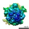 1524MC M: map data used to model this data C: citing same article ( |
|---|---|
| Similar structure data |


-Links
 PDBj
PDBj


- Assembly
| Deposited unit | 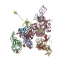
|
|---|---|
| 1 |
|
-Components
-RNA chain , 2 types, 2 molecules AB
| #1: RNA chain | Mass: 24890.121 Da / Num. of mol.: 1 / Source method: isolated from a natural source / Source: (natural) |
|---|---|
| #2: RNA chain | Mass: 24816.811 Da / Num. of mol.: 1 / Source method: isolated from a natural source / Source: (natural) |
-30S RNA helix ... , 2 types, 2 molecules EF
| #3: RNA chain | Mass: 5161.144 Da / Num. of mol.: 1 / Source method: isolated from a natural source / Source: (natural) |
|---|---|
| #4: RNA chain | Mass: 3827.336 Da / Num. of mol.: 1 / Source method: isolated from a natural source / Source: (natural) |
-50S RNA helix ... , 4 types, 4 molecules GIJK
| #5: RNA chain | Mass: 22580.502 Da / Num. of mol.: 1 / Source method: isolated from a natural source / Source: (natural) |
|---|---|
| #6: RNA chain | Mass: 9394.642 Da / Num. of mol.: 1 / Source method: isolated from a natural source / Source: (natural) |
| #7: RNA chain | Mass: 5765.451 Da / Num. of mol.: 1 / Source method: isolated from a natural source / Source: (natural) |
| #8: RNA chain | Mass: 4745.833 Da / Num. of mol.: 1 / Source method: isolated from a natural source / Source: (natural) |
-Protein , 3 types, 3 molecules CDH
| #9: Protein | Mass: 60476.660 Da / Num. of mol.: 1 / Fragment: EF4 Source method: isolated from a genetically manipulated source Source: (gene. exp.) |
|---|---|
| #10: Protein | Mass: 13636.961 Da / Num. of mol.: 1 / Source method: isolated from a natural source / Source: (natural) |
| #11: Protein | Mass: 14763.165 Da / Num. of mol.: 1 / Source method: isolated from a natural source / Source: (natural) |
-Experimental details
-Experiment
| Experiment | Method: ELECTRON MICROSCOPY |
|---|---|
| EM experiment | Aggregation state: PARTICLE / 3D reconstruction method: single particle reconstruction |
- Sample preparation
| Component | Name: Complex of elongating Escherichia coli 70S ribosome and EF4(LepA)-GMPPNP Type: COMPLEX |
|---|---|
| Buffer solution | Details: 20 mM HEPES-KOH (pH 7.6), 4.5 mM Mg(CH3COO)2, 150 mM NH4CH3COO, 4 mM B-mercaptoethanol, 2 mM spermidine, and 0.05 mM spermine |
| Specimen | Embedding applied: NO / Shadowing applied: NO / Staining applied: NO / Vitrification applied: YES |
| Vitrification | Instrument: FEI VITROBOT MARK I / Cryogen name: ETHANE |
- Electron microscopy imaging
| Experimental equipment |  Model: Tecnai Polara / Image courtesy: FEI Company |
|---|---|
| Microscopy | Model: FEI POLARA 300 |
| Electron gun | Electron source:  FIELD EMISSION GUN / Accelerating voltage: 300 kV / Illumination mode: FLOOD BEAM FIELD EMISSION GUN / Accelerating voltage: 300 kV / Illumination mode: FLOOD BEAM |
| Electron lens | Mode: BRIGHT FIELD / Nominal magnification: 39000 X / Nominal defocus max: 3900 nm / Nominal defocus min: 500 nm |
| Image recording | Electron dose: 20 e/Å2 / Film or detector model: KODAK SO-163 FILM |
- Processing
| EM software |
| ||||||||||||||||||||||||||||||||||||||||||
|---|---|---|---|---|---|---|---|---|---|---|---|---|---|---|---|---|---|---|---|---|---|---|---|---|---|---|---|---|---|---|---|---|---|---|---|---|---|---|---|---|---|---|---|
| Symmetry | Point symmetry: C1 (asymmetric) | ||||||||||||||||||||||||||||||||||||||||||
| 3D reconstruction | Resolution: 10.9 Å / Num. of particles: 41294 / Symmetry type: POINT | ||||||||||||||||||||||||||||||||||||||||||
| Atomic model building | Protocol: FLEXIBLE FIT Details: REFINEMENT PROTOCOL--rigid body and flexible fitting | ||||||||||||||||||||||||||||||||||||||||||
| Atomic model building |
| ||||||||||||||||||||||||||||||||||||||||||
| Refinement step | Cycle: LAST
|