 Movie
Movie Controller


+ Open data

- Basic information
| Entry | Database: EMDB / ID: EMD-2026 | |||||||||
|---|---|---|---|---|---|---|---|---|---|---|
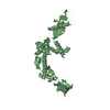
| Title | Structure of the proteasome subunit Rpn1 | |||||||||
 Map data Map data | Map of the proteasome subunit Rpn1 from S. cerevisiae | |||||||||
 Sample Sample |
| |||||||||
 Keywords Keywords | proteasome / rpn1 / 19S subunit | |||||||||
| Biological species |  | |||||||||
| Method | single particle reconstruction / negative staining / Resolution: 25.0 Å | |||||||||
 Authors Authors | He J / Kulkarni K / daFonseca P / Krutauz D / Glickman M / Barford D / Morris E | |||||||||
 Citation Citation | Journal: Structure / Year: 2012 Title: The structure of the 26S proteasome subunit Rpn2 reveals its PC repeat domain as a closed toroid of two concentric α-helical rings. Authors: Jun He / Kiran Kulkarni / Paula C A da Fonseca / Dasha Krutauz / Michael H Glickman / David Barford / Edward P Morris /  Abstract: The 26S proteasome proteolyses ubiquitylated proteins and is assembled from a 20S proteolytic core and two 19S regulatory particles (19S-RP). The 19S-RP scaffolding subunits Rpn1 and Rpn2 function to ...The 26S proteasome proteolyses ubiquitylated proteins and is assembled from a 20S proteolytic core and two 19S regulatory particles (19S-RP). The 19S-RP scaffolding subunits Rpn1 and Rpn2 function to engage ubiquitin receptors. Rpn1 and Rpn2 are characterized by eleven tandem copies of a 35-40 amino acid repeat motif termed the proteasome/cyclosome (PC) repeat. Here, we reveal that the eleven PC repeats of Rpn2 form a closed toroidal structure incorporating two concentric rings of α helices encircling two axial α helices. A rod-like N-terminal domain consisting of 17 stacked α helices and a globular C-terminal domain emerge from one face of the toroid. Rpn13, an ubiquitin receptor, binds to the C-terminal 20 residues of Rpn2. Rpn1 adopts a similar conformation to Rpn2 but differs in the orientation of its rod-like N-terminal domain. These findings have implications for understanding how 19S-RPs recognize, unfold, and deliver ubiquitylated substrates to the 20S core. | |||||||||
| History |
|
- Structure visualization
| Movie |
 Movie viewer Movie viewer |
|---|---|
| Structure viewer | EM map: SurfViewMolmilJmol/JSmol |
| Supplemental images |

- Downloads & links
-EMDB archive
| Map data | emd_2026.map.gz | 1.2 MB | EMDB map data format | |
|---|---|---|---|---|
| Header (meta data) | emd-2026-v30.xmlemd-2026.xml | 7.9 KB 7.9 KB | Display Display | EMDB header |
| Images | 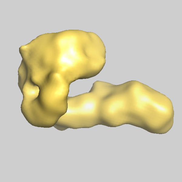 emd-2026.png emd-2026.png | 115.4 KB | ||
| Archive directory |  http://ftp.pdbj.org/pub/emdb/structures/EMD-2026ftp://ftp.pdbj.org/pub/emdb/structures/EMD-2026 http://ftp.pdbj.org/pub/emdb/structures/EMD-2026ftp://ftp.pdbj.org/pub/emdb/structures/EMD-2026 | HTTPS FTP |
-Validation report
| Summary document | emd_2026_validation.pdf.gz | 179.6 KB | Display | EMDB validaton report |
|---|---|---|---|---|
| Full document | emd_2026_full_validation.pdf.gz | 178.7 KB | Display | |
| Data in XML | emd_2026_validation.xml.gz | 5.6 KB | Display | |
| Arichive directory | https://ftp.pdbj.org/pub/emdb/validation_reports/EMD-2026ftp://ftp.pdbj.org/pub/emdb/validation_reports/EMD-2026 | HTTPS FTP |
-Related structure data

-Links
| EMDB pages | EMDB (EBI/PDBe) / EMDataResource |
|---|
-Map
| File | Download / File: emd_2026.map.gz / Format: CCP4 / Size: 7.8 MB / Type: IMAGE STORED AS FLOATING POINT NUMBER (4 BYTES) | ||||||||||||||||||||||||||||||||||||||||||||||||||||||||||||
|---|---|---|---|---|---|---|---|---|---|---|---|---|---|---|---|---|---|---|---|---|---|---|---|---|---|---|---|---|---|---|---|---|---|---|---|---|---|---|---|---|---|---|---|---|---|---|---|---|---|---|---|---|---|---|---|---|---|---|---|---|---|
| Annotation | Map of the proteasome subunit Rpn1 from S. cerevisiae | ||||||||||||||||||||||||||||||||||||||||||||||||||||||||||||
| Voxel size | X=Y=Z: 2.173 Å | ||||||||||||||||||||||||||||||||||||||||||||||||||||||||||||
| Density |
| ||||||||||||||||||||||||||||||||||||||||||||||||||||||||||||
| Symmetry | Space group: 1 | ||||||||||||||||||||||||||||||||||||||||||||||||||||||||||||
| Details | EMDB XML:
CCP4 map header:
| ||||||||||||||||||||||||||||||||||||||||||||||||||||||||||||
-Supplemental data
- Sample components
-Entire : Rpn1
| Entire | Name: Rpn1 |
|---|---|
| Components |
|
-Supramolecule #1000: Rpn1
| Supramolecule | Name: Rpn1 / type: sample / ID: 1000 / Number unique components: 1 |
|---|---|
| Molecular weight | Theoretical: 109.492 KDa |
-Macromolecule #1: Rpn1
| Macromolecule | Name: Rpn1 / type: protein_or_peptide / ID: 1 / Name.synonym: Rpn1 / Number of copies: 1 / Oligomeric state: Monomer / Recombinant expression: Yes |
|---|---|
| Source (natural) | Organism: |
| Molecular weight | Theoretical: 109.492 KDa |
| Recombinant expression | Organism:  |
-Experimental details
-Structure determination
| Method | negative staining |
|---|---|
 Processing Processing | single particle reconstruction |
| Aggregation state | particle |
-Sample preparation
| Concentration | 0.1 mg/mL |
|---|---|
| Staining | Type: NEGATIVE / Details: 2% uranyl acetate |
| Grid | Details: Carbon film on quantifoil |
| Vitrification | Cryogen name: NONE / Instrument: OTHER |
- Electron microscopy
| Microscope | FEI TECNAI F20 |
|---|---|
| Image recording | Category: CCD / Film or detector model: TVIPS TEMCAM-F415 (4k x 4k) / Number real images: 71 / Average electron dose: 100 e/Å2 / Bits/pixel: 16 |
| Electron beam | Acceleration voltage: 200 kV / Electron source:  FIELD EMISSION GUN FIELD EMISSION GUN |
| Electron optics | Illumination mode: FLOOD BEAM / Imaging mode: BRIGHT FIELD / Nominal defocus max: 0.9 µm / Nominal defocus min: 0.8 µm / Nominal magnification: 80000 |
| Sample stage | Specimen holder: Eucentric / Specimen holder model: SIDE ENTRY, EUCENTRIC |
| Experimental equipment |  Model: Tecnai F20 / Image courtesy: FEI Company |
-Image processing
| Final reconstruction | Applied symmetry - Point group: C1 (asymmetric) / Resolution.type: BY AUTHOR / Resolution: 25.0 Å / Resolution method: FSC 0.5 CUT-OFF / Software - Name: Imagic |
|---|
