 Movie
Movie Controller


+ Open data

- Basic information
| Entry | Database: PDB / ID: 4c8q | ||||||
|---|---|---|---|---|---|---|---|
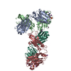
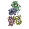
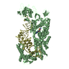
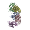
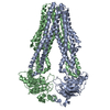
| Title | Crystal structure of the yeast Lsm1-7-Pat1 complex | ||||||
 Components Components |
| ||||||
 Keywords Keywords | TRANSCRIPTION / MRNA DECAPPING / SM FOLD / MRNA DEGRADATION | ||||||
| Function / homology |  Function and homology information Function and homology informationmRNA decay by 5' to 3' exoribonuclease / Lsm1-7-Pat1 complex / U6 snRNP / deadenylation-dependent decapping of nuclear-transcribed mRNA / formation of translation preinitiation complex / sno(s)RNA-containing ribonucleoprotein complex / spliceosomal tri-snRNP complex / P-body assembly / regulation of translational initiation / U2-type prespliceosome ...mRNA decay by 5' to 3' exoribonuclease / Lsm1-7-Pat1 complex / U6 snRNP / deadenylation-dependent decapping of nuclear-transcribed mRNA / formation of translation preinitiation complex / sno(s)RNA-containing ribonucleoprotein complex / spliceosomal tri-snRNP complex / P-body assembly / regulation of translational initiation / U2-type prespliceosome / tRNA processing / nuclear-transcribed mRNA catabolic process, deadenylation-dependent decay / precatalytic spliceosome / nuclear-transcribed mRNA catabolic process / U6 snRNA binding / cellular response to glucose starvation / U4/U6 x U5 tri-snRNP complex / spliceosomal snRNP assembly / catalytic step 2 spliceosome / negative regulation of translational initiation / spliceosomal complex / maturation of SSU-rRNA / mRNA splicing, via spliceosome / P-body / kinetochore / mRNA processing / cytoplasmic stress granule / rRNA processing / cytosolic small ribosomal subunit / ribonucleoprotein complex / cell division / mRNA binding / chromatin binding / nucleolus / RNA binding / nucleus / cytoplasm Similarity search - Function | ||||||
| Biological species |  | ||||||
| Method |  X-RAY DIFFRACTION / SYNCHROTRON / MOLECULAR REPLACEMENT / Resolution: 3.698 Å X-RAY DIFFRACTION / SYNCHROTRON / MOLECULAR REPLACEMENT / Resolution: 3.698 Å | ||||||
 Authors Authors | Sharif, H. / Conti, E. | ||||||
 Citation Citation | Journal: Cell Rep. / Year: 2013 Title: Architecture of the Lsm1-7-Pat1 Complex: A Conserved Assembly in Eukaryotic Mrna Turnover Authors: Sharif, H. / Conti, E. | ||||||
| History |
| ||||||
| Remark 650 | HELIX DETERMINATION METHOD: AUTHOR PROVIDED. |
- Structure visualization
| Structure viewer | Molecule: MolmilJmol/JSmol |
|---|
- Downloads & links
-Download
| PDBx/mmCIF format | 4c8q.cif.gz | 181.6 KB | Display | PDBx/mmCIF format |
|---|---|---|---|---|
| PDB format | pdb4c8q.ent.gz | 136.9 KB | Display | PDB format |
| PDBx/mmJSON format | 4c8q.json.gz | Tree view | PDBx/mmJSON format | |
| Others |  Other downloads Other downloads |
-Validation report
| Arichive directory | https://data.pdbj.org/pub/pdb/validation_reports/c8/4c8qftp://data.pdbj.org/pub/pdb/validation_reports/c8/4c8q | HTTPS FTP |
|---|
-Related structure data
| Related structure data | 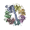 4c92SC S: Starting model for refinement C: citing same article ( |
|---|---|
| Similar structure data |


-Links
 PDBj
PDBj


- Assembly
| Deposited unit | 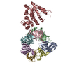
| ||||||||
|---|---|---|---|---|---|---|---|---|---|
| 1 |
| ||||||||
| Unit cell |
|
-Components
-Protein , 2 types, 2 molecules AH
| #1: Protein | Mass: 11935.651 Da / Num. of mol.: 1 / Fragment: RESIDUES 45-143 Source method: isolated from a genetically manipulated source Source: (gene. exp.) Strain: S288C / Production host:  |
|---|---|
| #8: Protein | Mass: 37934.699 Da / Num. of mol.: 1 / Fragment: RESIDUES 456-783 Source method: isolated from a genetically manipulated source Source: (gene. exp.) Strain: S288C / Production host: |
-U6 SNRNA-ASSOCIATED SM-LIKE PROTEIN ... , 6 types, 6 molecules BCDEFG
| #2: Protein | Mass: 12216.880 Da / Num. of mol.: 1 / Fragment: RESIDUES 2-95 Source method: isolated from a genetically manipulated source Source: (gene. exp.) Strain: S288C / Production host: |
|---|---|
| #3: Protein | Mass: 10039.262 Da / Num. of mol.: 1 / Fragment: RESIDUES 1-89 Source method: isolated from a genetically manipulated source Source: (gene. exp.) Strain: S288C / Production host: |
| #4: Protein | Mass: 13035.543 Da / Num. of mol.: 1 / Fragment: RESIDUES 1-114 Source method: isolated from a genetically manipulated source Source: (gene. exp.) Strain: S288C / Production host: |
| #5: Protein | Mass: 10432.954 Da / Num. of mol.: 1 / Fragment: RESIDUES 1-93 Source method: isolated from a genetically manipulated source Source: (gene. exp.) Strain: S288C / Production host: |
| #6: Protein | Mass: 9406.579 Da / Num. of mol.: 1 / Fragment: RESIDUES 1-86 Source method: isolated from a genetically manipulated source Source: (gene. exp.) Strain: S288C / Production host: |
| #7: Protein | Mass: 13027.045 Da / Num. of mol.: 1 / Fragment: RESIDUES 1-115 Source method: isolated from a genetically manipulated source Source: (gene. exp.) Strain: S288C / Production host: |
-Non-polymers , 1 types, 2 molecules 
| #9: Chemical |  Mass: 58.933 Da / Num. of mol.: 2 / Source method: obtained synthetically / Formula: Co Mass: 58.933 Da / Num. of mol.: 2 / Source method: obtained synthetically / Formula: Co |
|---|
-Experimental details
-Experiment
| Experiment | Method: X-RAY DIFFRACTION |
|---|
- Sample preparation
| Crystal | Density Matthews: 3.89 Å3/Da / Density % sol: 68.39 % Description: CHIMERIC MODEL WAS MADE FOR MOLECULAR REPLACEMENT |
|---|---|
| Crystal grow | Details: 100 MM HEPES PH 7.0, 20% MPD, 10 MM HEXAMINE COBALT(III) CHLORIDE |
-Data collection
| Diffraction | Mean temperature: 100 K |
|---|---|
| Diffraction source | Source: SYNCHROTRON / Site: ESRF  / Beamline: ID23-2 / Wavelength: 0.8729 / Beamline: ID23-2 / Wavelength: 0.8729 |
| Detector | Type: MARMOSAIC 225 mm CCD / Detector: CCD / Date: Aug 1, 2013 |
| Radiation | Protocol: SINGLE WAVELENGTH / Monochromatic (M) / Laue (L): M / Scattering type: x-ray |
| Radiation wavelength | Wavelength: 0.8729 Å / Relative weight: 1 |
| Reflection | Resolution: 3.7→84.68 Å / Num. obs: 36441 / % possible obs: 99.4 % / Observed criterion σ(I): 2 / Redundancy: 1.8 % / Biso Wilson estimate: 133.3 Å2 / Rmerge(I) obs: 0.07 / Net I/σ(I): 11.3 |
| Reflection shell | Resolution: 3.7→3.9 Å / Redundancy: 1.6 % / Rmerge(I) obs: 0.69 / Mean I/σ(I) obs: 1.4 / % possible all: 97.6 |
- Processing
| Software |
| ||||||||||||||||||||||||||||||||||||||||||||||||||||||||||||||||||||||||||||||||||||||||||||||||||
|---|---|---|---|---|---|---|---|---|---|---|---|---|---|---|---|---|---|---|---|---|---|---|---|---|---|---|---|---|---|---|---|---|---|---|---|---|---|---|---|---|---|---|---|---|---|---|---|---|---|---|---|---|---|---|---|---|---|---|---|---|---|---|---|---|---|---|---|---|---|---|---|---|---|---|---|---|---|---|---|---|---|---|---|---|---|---|---|---|---|---|---|---|---|---|---|---|---|---|---|
| Refinement | Method to determine structure: MOLECULAR REPLACEMENT Starting model: PDB ENTRY 4C92 Resolution: 3.698→43.42 Å / SU ML: 0.67 / σ(F): 39.43 / Phase error: 31.14 / Stereochemistry target values: ML
| ||||||||||||||||||||||||||||||||||||||||||||||||||||||||||||||||||||||||||||||||||||||||||||||||||
| Solvent computation | Shrinkage radii: 0.9 Å / VDW probe radii: 1.11 Å / Solvent model: FLAT BULK SOLVENT MODEL | ||||||||||||||||||||||||||||||||||||||||||||||||||||||||||||||||||||||||||||||||||||||||||||||||||
| Displacement parameters | Biso mean: 129.27 Å2 | ||||||||||||||||||||||||||||||||||||||||||||||||||||||||||||||||||||||||||||||||||||||||||||||||||
| Refinement step | Cycle: LAST / Resolution: 3.698→43.42 Å
| ||||||||||||||||||||||||||||||||||||||||||||||||||||||||||||||||||||||||||||||||||||||||||||||||||
| Refine LS restraints |
| ||||||||||||||||||||||||||||||||||||||||||||||||||||||||||||||||||||||||||||||||||||||||||||||||||
| LS refinement shell |
|
