National Institutes of Health/National Institute of Diabetes and Digestive and Kidney Disease
DK027044
United States
Citation
Journal: Nat Commun / Year: 2019 Title: Structural basis for the clamping and Ca activation of SNARE-mediated fusion by synaptotagmin. Authors: Kirill Grushin / Jing Wang / Jeff Coleman / James E Rothman / Charles V Sindelar / Shyam S Krishnakumar / Abstract: Synapotagmin-1 (Syt1) interacts with both SNARE proteins and lipid membranes to synchronize neurotransmitter release to calcium (Ca) influx. Here we report the cryo-electron microscopy structure of ...Synapotagmin-1 (Syt1) interacts with both SNARE proteins and lipid membranes to synchronize neurotransmitter release to calcium (Ca) influx. Here we report the cryo-electron microscopy structure of the Syt1-SNARE complex on anionic-lipid containing membranes. Under resting conditions, the Syt1 C2 domains bind the membrane with a magnesium (Mg)-mediated partial insertion of the aliphatic loops, alongside weak interactions with the anionic lipid headgroups. The C2B domain concurrently interacts the SNARE bundle via the 'primary' interface and is positioned between the SNAREpins and the membrane. In this configuration, Syt1 is projected to sterically delay the complete assembly of the associated SNAREpins and thus, contribute to clamping fusion. This Syt1-SNARE organization is disrupted upon Ca-influx as Syt1 reorients into the membrane, likely displacing the attached SNAREpins and reversing the fusion clamp. We thus conclude that the cation (Mg/Ca) dependent membrane interaction is a key determinant of the dual clamp/activator function of Synaptotagmin-1.
History
Deposition
Oct 17, 2018
-
Header (metadata) release
Nov 14, 2018
-
Map release
Apr 24, 2019
-
Update
Nov 6, 2019
-
Current status
Nov 6, 2019
Processing site: RCSB / Status: Released
-
Structure visualization
Movie
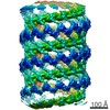
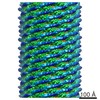
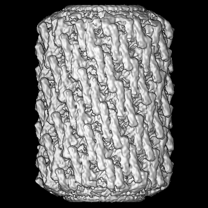
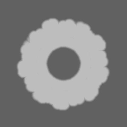
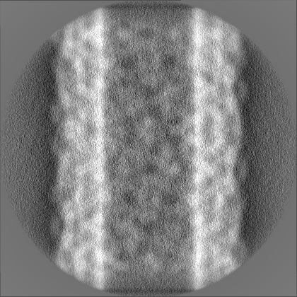
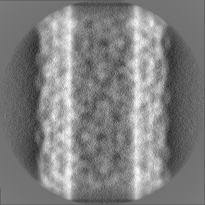
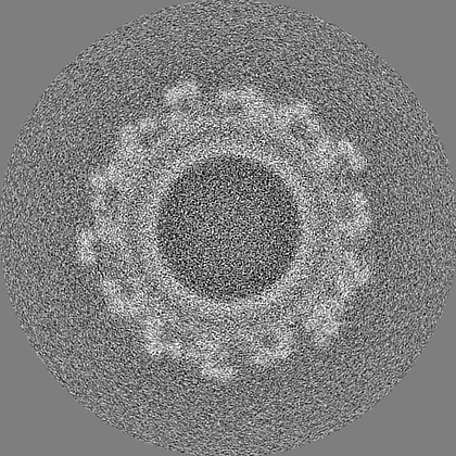
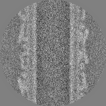
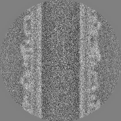
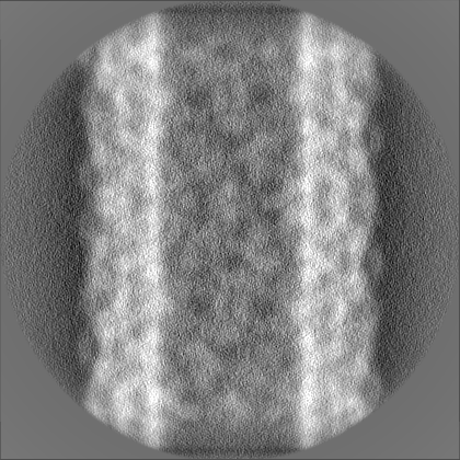
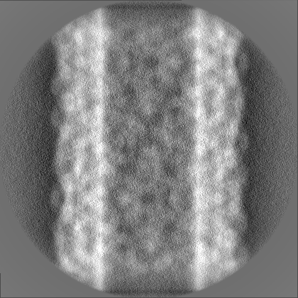
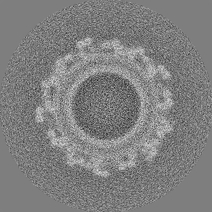
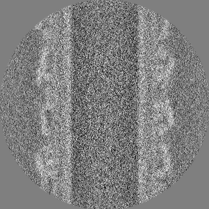
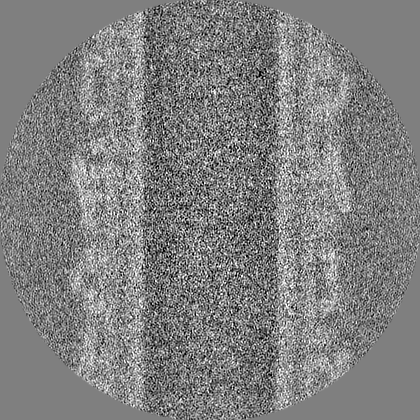
Surface view with section colored by density value
Entire : Synaptotagmin 1 C2AB in a complex with the SNAREpin assembly boun...
Entire
Name: Synaptotagmin 1 C2AB in a complex with the SNAREpin assembly bound to the negatively charged phospholipid nanotube in presence of Mg2+.
Components
Complex: Synaptotagmin 1 C2AB in a complex with the SNAREpin assembly bound to the negatively charged phospholipid nanotube in presence of Mg2+.
Protein or peptide: Synaptotagmin 1
Protein or peptide: Vesicle-associated membrane protein 2
Protein or peptide: Syntaxin-1A
Protein or peptide: Synaptosomal-associated protein 25
Protein or peptide: Synaptosomal-associated protein 25
-
Supramolecule #1: Synaptotagmin 1 C2AB in a complex with the SNAREpin assembly boun...
Supramolecule
Name: Synaptotagmin 1 C2AB in a complex with the SNAREpin assembly bound to the negatively charged phospholipid nanotube in presence of Mg2+. type: complex / ID: 1 / Parent: 0 / Macromolecule list: all
Type of model: INSILICO MODEL / In silico model: Featureless hollow cylinder
Final angle assignment
Type: NOT APPLICABLE
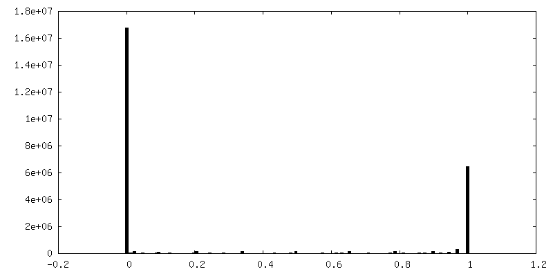
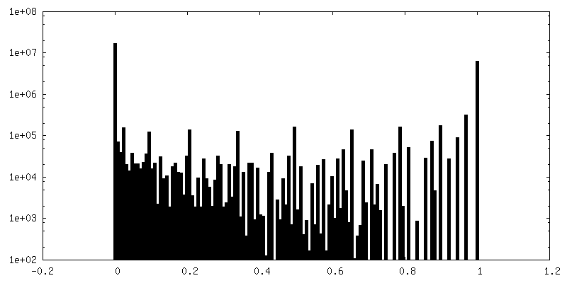
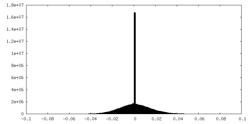
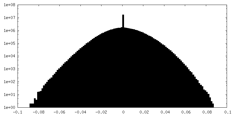
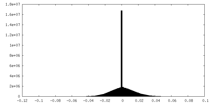
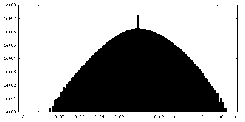
FSC plot (resolution estimation)
-
Atomic model buiding 1
Refinement
Protocol: RIGID BODY FIT
Output model
PDB-6mti: Synaptotagmin-1 C2A, C2B domains and SNARE-pin proteins (5CCI) individually docked into Cryo-EM map of C2AB-SNARE complexes helically organized on lipid nanotube surface in presence of Mg2+
+
About Yorodumi
-
News
-
Feb 9, 2022. New format data for meta-information of EMDB entries
New format data for meta-information of EMDB entries
Version 3 of the EMDB header file is now the official format.
The previous official version 1.9 will be removed from the archive.
In the structure databanks used in Yorodumi, some data are registered as the other names, "COVID-19 virus" and "2019-nCoV". Here are the details of the virus and the list of structure data.
Jan 31, 2019. EMDB accession codes are about to change! (news from PDBe EMDB page)
EMDB accession codes are about to change! (news from PDBe EMDB page)
The allocation of 4 digits for EMDB accession codes will soon come to an end. Whilst these codes will remain in use, new EMDB accession codes will include an additional digit and will expand incrementally as the available range of codes is exhausted. The current 4-digit format prefixed with “EMD-” (i.e. EMD-XXXX) will advance to a 5-digit format (i.e. EMD-XXXXX), and so on. It is currently estimated that the 4-digit codes will be depleted around Spring 2019, at which point the 5-digit format will come into force.
The EM Navigator/Yorodumi systems omit the EMD- prefix.
Related info.:Q: What is EMD? / ID/Accession-code notation in Yorodumi/EM Navigator
Yorodumi is a browser for structure data from EMDB, PDB, SASBDB, etc.
This page is also the successor to EM Navigator detail page, and also detail information page/front-end page for Omokage search.
The word "yorodu" (or yorozu) is an old Japanese word meaning "ten thousand". "mi" (miru) is to see.
Related info.:EMDB / PDB / SASBDB / Comparison of 3 databanks / Yorodumi Search / Aug 31, 2016. New EM Navigator & Yorodumi / Yorodumi Papers / Jmol/JSmol / Function and homology information / Changes in new EM Navigator and Yorodumi
 Movie
Movie Controller

 Yorodumi
 Open data
 Basic information
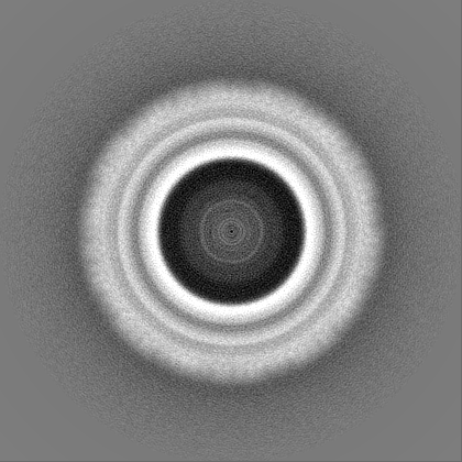
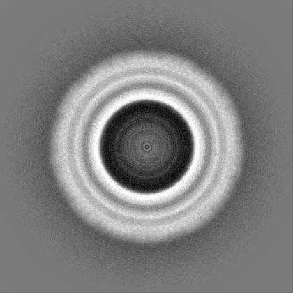
 Map data
 Sample
 Function and homology information
 Authors
 United States, 1 items
  Citation
 Structure visualization
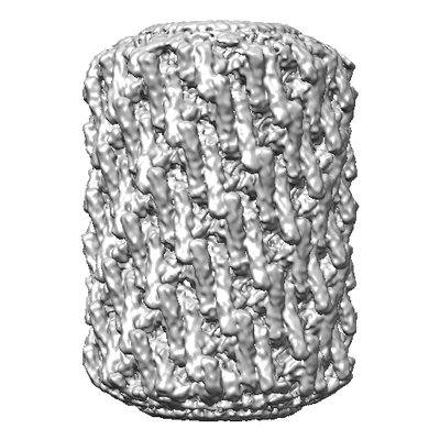
 Downloads & links
 emd_9231.png
 http://ftp.pdbj.org/pub/emdb/structures/EMD-9231


 Z (Sec.)
 Y (Row.)
 X (Col.)


 Sample components
 Processing
 Electron microscopy
 FIELD EMISSION GUN

